A mass growing in the abdomen of a 17-year-old-girl in Patna, India was so huge, it was filling and distorting her entire abdominal cavity. But instead of a garden-variety tumour, doctors discovered a mass that contained teeth, hair, bones, fat and cartilage.
There is a type of tumour that can include these types of tissue, called a teratoma. But this was no teratoma. It seemed to be something much rarer - possibly the girl's own twin, absorbed while they were both still in the uterus.
It's a condition called foetus in foetu (FIF), and it's not usually seen in anyone past the age of 15.
According to the case report, the patient sought medical attention, as for the past five years she had noticed a lump growing in her abdomen. Sometimes it caused her pain; sometimes, when eating, she felt full really quickly.
On examination, the doctors felt a large, hard mass, irregularly shaped. Blood tests came back normal, so they performed a scan.
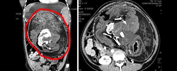
"An abdominal contrast-enhanced computed tomography (CECT) scan showed a well-defined mass that measured approximately 25 by 23 by 15 centimetres (9.8 by 9.1 by 5.9 inches), extending from epigastrium [just above the stomach] to upper pelvis," they wrote in their case report.
"It was showing fat density areas, soft tissue and multiple calcified density components of various sizes and shapes resembling the shape of vertebrae, ribs and long bones. This mass was causing displacement and compression of adjacent abdominal viscera."
The presence of fat, vertebrae and ribs, as well as the duration of the complaint and the size of the mass, led doctors to diagnose the teenage patient with FIF. They scheduled surgery to remove the mass.
Sure enough, what the doctors removed from the girl's abdomen seemed to be a partially formed human. The team described a mass made from "hairy cheesy material, multiple teeth and structures resembling limb buds," including fat, cartilage and bone, as well as neural, intestinal and skin tissue.
It's not actually known how FIF develops. The parasitic twin theory, wherein one twin of an identical foetal pair is absorbed by the other, is one. This is supported by the affected demographic: usually young children, as the condition is typically detected in newborns. The bizarre phenomenon is thought to occur in just 1 in 500,000 live births, and the parasitic mass doesn't usually survive for long.
Another is that it may be a particularly well developed form of teratoma, which may be how it occurs in older patients. Previously, only seven other cases of FIF had been reported in patients over the age of 15.
It is difficult to tell the difference between the two, the doctors noted. One key difference between FIF and teratoma is that the diagnostic criteria for the former include a vertebral axis with organs forming, while the latter is a disorganised mass of pluripotent cells.
It could be possible that there is a type of teratoma that is more developed than just a wobbly bit of flesh jelly with teeth and hair and the occasional eye. Given, however, that this patient's parasite had spinal bones and ribs, the official diagnosis is foetus in foetu.
The surgery was a success, with most of the mass removed. Some, however, had to be left behind - it had adhered too closely to the blood vessels of the gastrointestinal tract, and removing it would have risked significant blood loss.
The patient is doing well, but will have to return for annual check-ups to make sure that the remaining tissue hasn't turned cancerous.
"I was much worried about my abdominal lump, after operation I am feeling very well and my abdomen is now flat and my parents are also very happy," she stated in the case report. "Thanks to all operating doctors."
The report has been published in BMJ Case Reports.