A group of five children in China have been given new ears - based on detailed 3D models and grown from their own cells - in a world first for this kind of treatment.
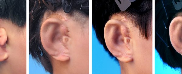
The kids, aged between 6 and 9, all had microtia, where the external part of the ear ends up deformed. In these cases the condition was unilateral, affecting only one side, so scientists were able to create high-resolution scans of their healthy ears to help grow replacement ones.
Now the team of tissue engineers and plastic surgeons has proved these techniques can work in human beings, they could offer a new lease of life for people living with microtia or other similar conditions.
"The results represent a significant breakthrough in clinical translation of tissue engineered human ear-shaped cartilage given the established in vitro engineering technique and suitable surgical procedure," write the researchers in their published paper.
 (EBioMedicine)
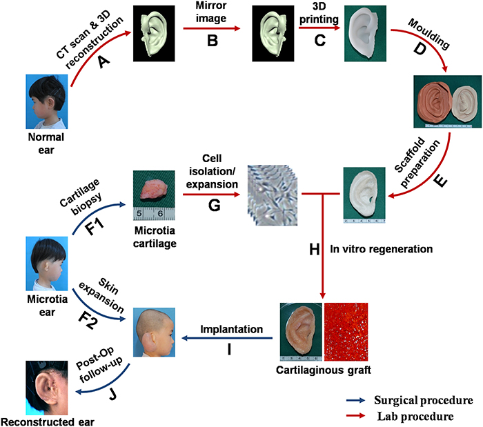
Cartilage cells called chondrocytes were harvested from the non-deformed ears by the scientists and then used to create a new ear for the other side of the head through a process of cell culturing.
With the help of computed tomography or CT scans of the healthy ears, a 3D-printed framework was created that the newly growing ear could expand into. Over time, natural cells replaced almost all of the artificial scaffolding.
Finally, the new ears were attached and reconstruction was completed, with some small cosmetic surgery procedures applied afterwards.
This kind of biological technology is actually several years old, but this is the first time it's been used so effectively in human beings – the first of these implants was fitted 30 months ago, suggesting the long-term prospects are good.
"The delivery of shaped cartilage for the reconstruction of microtia has been a goal of the tissue engineering community for more than two decades," Lawrence Bonassar, a biomedical engineering professor from Cornell University in New York who wasn't involved in the study, told Jacqueline Howard at CNN.
"This work clearly shows tissue engineering approaches for reconstruction of the ear and other cartilaginous tissues will become a clinical reality very soon. The aesthetics of the tissue produced are on par with what can be expected of the best clinical procedures at the present time."
Microtia rates are as high as 17.4 in 10,000 in some countries, affecting both hearing and self-confidence of the kids who are born with it.
Current treatments include fitting an artificial ear frame, or creating a new ear from rib cartilage, but this new approach beats them all in terms of both appearance and lessening the damage on the patient's body.
"Surgeons have been toying with the idea of removing cartilage tissue from a patient and distilling that tissue into individual cellular components and then expanding those cellular components," Tessa Hadlock from the Massachusetts Eye and Ear Infirmary, who wasn't involved in the study, told CNN.
"The thing that is novel about this is that for the first time, they have done it in a series of five patients, and they have good long-term follow-up that shows the results of the ears that were grown from that harvested cartilage."
However, there are some caveats to note – 2.5 years is a good stretch but the artificial parts of the ear haven't yet fully degraded, so further monitoring up to 5 years is going to be needed before we're sure this is a success.
What's more, two of the cases showed slight distortions in the growth of the ears, which scientists will have to carefully monitor.
Nevertheless it's a promising step forward for these procedures, as well as a potentially life-changing new option for those with microtia, if it becomes widely available.
"We were able to successfully design, fabricate, and regenerate patient-specific external ears," write the researchers. "Further efforts remain necessary to eventually translate this prototype work into routine clinical practices."
The research has been published in EBioMedicine.