Scientists in Canada have developed a new method of growing human tissue outside the body, creating a miniature lattice structure that's capable of providing an external matrix for living cells.
Called AngioChip, the researchers say their 'person on a chip' technology could be a new platform for testing the effects of drugs on human tissue, with the mini 3D scaffold constituting a more realistic environment for growing cells than the flat layout of a petri dish.
"It's a fully three-dimensional structure complete with internal blood vessels," said chemical engineer Milica Radisic from the University of Toronto. "It behaves just like vasculature, and around it there is a lattice for other cells to attach and grow."
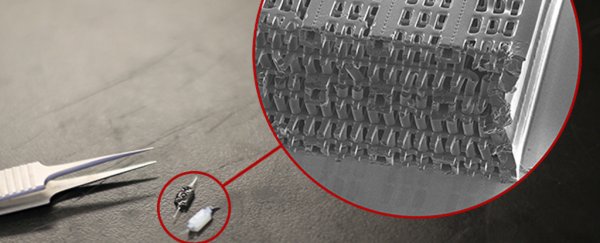
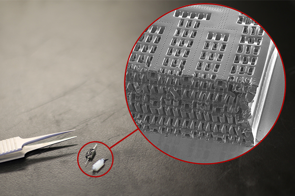
Built from a biodegradable and biocompatible polymer called POMaC, the miniature scaffold is constructed from a series of thin layers that resemble microchips, each indented with a pattern of tiny channels measuring between 50 to 100 micrometres wide (about the same diameter as a human hair).
Once stacked and bonded together via UV light, these layers become a 3D structure of synthetic blood vessels. The lattice network is then bathed in a liquid containing living cells. The cells attach to the structure, and begin growing inside and outside of the tiny channels stamped in the polymer.
"Previously, people could only do this using devices that squish the cells between sheets of silicone and glass," said Radisic. "You needed several pumps and vacuum lines to run just one chip. Our system runs in a normal cell culture dish, and there are no pumps; we use pressure heads to perfuse media through the vasculature. The wells are open, so you can easily access the tissue."
 Tyler Irving / Boyang Zhang / Kevin Soobrian
The engineers have so far used AngioChip to build small-scale living models of heart and liver tissues that function just like real organs. "Our liver actually produced urea and metabolised drugs," said Radisic.
What's more, connecting the blood vessels of two different artificial organs on AngioChips lets the researchers study the interactions between them, providing a testbed at the organ level.
Such testing could more accurately detect dangerous side effects of potential or existing medications, or safely study related interactions between various human organ compartments.
"In the last few years, it has become possible to order cultures of human cells for testing, but they're grown on a plate, a two-dimensional environment," said Radisic. "They don't capture all the functional hallmarks of a real heart muscle, for example."
The researchers' technology is reported in Nature Materials. They are now seeking to commercialise the system, which could end up having applications even beyond drug testing. They say it's possible the artificial AngioChip organs could be grown into tissues for directly implanting into patients' bodies to repair damaged organs.
So far, the researchers have only tested this application in rats, but the advantage of the system is that the polymer scaffold is itself biodegradable, so after time it safely dissipates within the subject's body, leaving only the newly grown tissue.
It sounds like it'll be a while before we see this kind of technique used for human patients, but even so, we can't wait to see where this research might lead.