Using a mix of electrical stimulation and intense physical therapy, nine people with chronic spinal injuries have had their ability to walk restored.
All suffered from severe or complete paralysis as a result of damage to their spinal cord. Incredibly, the volunteers all saw improvements immediately, and continued to show improvements five months later.
A recent study by researchers from the Swiss research group NeuroRestore has identified the exact nerve groups stimulated by the therapy, using mice as a starting point.
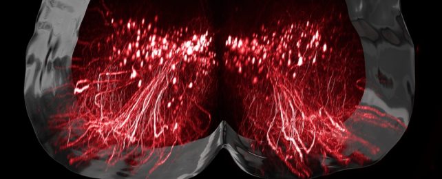
The nerve cells that orchestrate walking are found in the section of spinal cord running through our lower backs. Injuries to our spinal cord can interrupt the chain of signals from the brain, preventing us from walking even when these specific lumbar neurons are still intact.
Unable to receive commands, these 'walking' neurons effectively become nonfunctional, potentially leading to a permanent paralysis of the legs.
Previous research demonstrated electrical stimulation of the spinal cord can reverse such paralysis, but how this occurred wasn't clear. So neuroscientist Claudia Kathe from the Swiss Federal Institute of Technology Lausanne (EPFL) and colleagues tested a technology called epidural electrical stimulation in nine individuals, as well as in an animal model.
The spinal cord was stimulated by a surgically implanted neurotransmitter. Meanwhile, patients also underwent a process of intensive neurorehabilitation that involved a robotic support system assisting them while they moved in multiple directions.
The patients went through five months of stimulation and rehabilitation, four to five times per week. Amazingly, all of the volunteers were then able to take steps with the aid of a walker.
To the researchers' surprise, the recovered patients actually showed a reduction in neural activity in the lumbar spinal cord during walking. The team believes this is due to the activity being refined to a specific subset of neurons that are essential for walking.
"When you think about it, it should not be a surprise," Courtine told Dyani Lewis at Nature, "because in the brain, when you learn a task, that's exactly what you see – there are less and less neurons activated" as you get better at it.
So Kathe and team modeled the process in mice and used a combination of RNA sequencing and spatial transcriptomics – a technique that allows scientists to measure and map gene activity in specific tissues – to understand which cells were doing what.
They identified a single population of previously unknown neurons that can step up to take over after an injury, found within the intermediate laminae of the lumbar spinal cord.
This tissue, made up of cells called SCVsx2::Hoxa10 neurons, don't appear to be needed for walking in healthy animals, but they seem to be essential for recovering after a spinal injury, as destroying them prevented mice from recovering. Their recruitment is, however, activity dependent.
SCVsx2::Hoxa10 neurons are "uniquely positioned" to transform information from the brainstem into executive commands. These are then broadcast to the neurons that are responsible for the production of walking, Kathe and colleagues explain in their paper.
This is only one component of a very complicated chain of messaging and receiving cells, so there's still a lot that remains to be investigated.
But, "these experiments confirmed that the participation of SCVsx2::Hoxa10 neurons is a fundamental requirement for the recovery of walking after paralysis," the researchers concluded.
This new understanding could in time lead to more treatment options, and may provide a better quality of life for people with all sorts of other spinal cord injuries too.
Their research was published in Nature.