Want to see something that makes scientists gasp? Feast your eyes on this video, showing a direct observation of CRISPR-Cas9 munching up a piece of DNA. In real-time.
It's behaving exactly how scientists predicted CRISPR edits DNA, but this grainy video is the first time a direct observation of the process has been made.
The video was shown to a group of scientists at the CRISPR 2017 conference in Big Sky, Montana in June, by structural biologist Osamu Nureki of the University of Tokyo.
"I was sitting in the front, and I just heard this gasp from everyone behind me," biochemist Sam Sternberg told The Atlantic. Watch it yourself here:
Single-molecule movie of DNA search and cleavage by CRISPR-Cas9. pic.twitter.com/3NQxmbvzJF
— hnisimasu (@hnisimasu) November 10, 2017
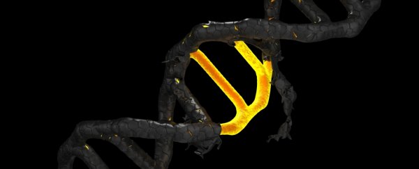
CRISPR-Cas9 has been making waves in recent years as a tool for editing genomes. It consists of a family of DNA sequences that forms part of the immune system of bacteria.
When CRISPR is filled with virus DNA, it copies it into short RNA sequences called 'guide RNA'. Cas enzymes will follow the guide RNA through cells, and when it finds DNA that matches the guide RNA, it destroys it by cleaving it - like a pair of molecular scissors.
In 2012, scientists demonstrated the potential of the CRISPR-Cas9 system. By intentionally encoding CRISPR with specific DNA, the system can be used for highly targeted genome editing.
It's been proven to work on many species, and so far has been used to treat a genetic disease in mice, change the colour of a flower, eliminate HIV in living animals, slow the growth of cancer cells, and remove a gene that causes heart disease from human embryos.
But directly observing CRISR in action had never been done until now. Scientists know it works, because they get results, but the mechanism whereby it works has, until now, been essentially hypothetical. What is happening is simply too small for normal imaging methods.
To get around this problem, Nureki and his team used a technique called high-speed atomic force microscopy. An atomic force microscope consists of a very sharp-tipped probe on the free end of a cantilever. The probe lowers towards the surface and deflects away, over and over.
As it does this, a laser detects the slight changes of the deflections of the cantilever as it moves over raised surface features. These are recorded to create an image of what the probe is scanning.
One of the researchers on the team, Hiroshi Nishimasu, posted the video to Twitter. The yellow blob is Cas9, the brown strands are DNA. It takes CRISPR-Cas9 an order of seconds to cut through the DNA strand once it has cleaved to it.
That grainy, blobby, 10-second video? It's one of the most amazing scientific images we've ever seen.
The research has been published in the journal Nature Communications.