
Cells come in all 3D shapes and sizes, but when we look at them through a microscope, we only see a 2D image.
That's okay for creating a basic image of the structures, but it's not enough when you want to dig deeper, because although two things in a cell might be far apart, when scientists look through a microscope in 2D, they can appear to be right on top of each other.
An international collaboration of researchers have been looking for a way to overcome this problem, and they think they've found one - using tiny mirrors to create a 3D view of cells.
The researchers, from the University of Technology Sydney (UTS) in Australia, Peking University in China, and the Georgia Institute of Technology in the US, say that although this technology is simple, it should allow for scientists to gather much better information about the third dimension of cells.
"This simple technology is allowing us to see the details of cells that have never been seen before," said one of the researchers, Dayong Jin from UTS.
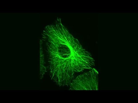
"A single cell is about 10 micrometres; inside that is a nuclear core about 5 micrometres, and inside that are tiny holes, called the 'nuclear pore complex' that as a gate regulates the messenger bio-molecules, but measures between one-50th and one-20th of a micrometre. With this super-resolution microscopy we are able to see the details of those tiny holes."
The new technology, dubbed MEANS (mirror-enhanced, axial-narrowing, super-resolution) microscopy, works by capturing layers of the cell at different focal points, giving the researchers the ability to see the changes in layers of the cell.
The process works by growing cells on small, custom-made mirrors. A glass cover slide is placed on top of the cells, and then the mirror can be added in place of a usual slide in a confocal or wide-field microscope.
"Most people are not growing cells on mirrors, so it required some work to get the cell culture conditions correct," said one of the UTS team, Phil Santangelo. "We had to make sure the mirror coating didn't affect cell growth, and staining the cells to make them fluoresce also required some adaption. Ultimately, growing cells on the mirrors became a simple process."
"Previously, the vision of biologists was blurred by the large axial and lateral resolution," says another researcher, Peng Xi from Peking University.
"This was like reading newspapers printed on transparent plastic; many layers were overlapped. By placing a mirror beneath the specimen, we can generate a narrowed focal spot so there is only one layer of the newspaper to read… every word becomes crystal clear."
Already the researchers have been able to investigate the ring structure of the nuclear pore complex using the new technology, as well as the structure of human respiratory syncytial virus.
The development could have wide reaching implications for the microscopy field, and could help scientists to see cells from a whole new perspective, and in more detail than ever before.
We're looking forward to seeing how this progresses.
The research was published this month in Light: Science & Applications.
UTS Science is a sponsor of ScienceAlert. Find out more about their research.