A new device, which can sort and isolate individual cells based on their ability to move around the body, could help us prevent the spread of cancer, researchers say.
Cancer occurs after a single cell in a tissue is genetically damaged and begins to multiply uncontrollably, eventually leading to the formation of a tumour. The disease gets particularly deadly when it metastasises, or spreads to other parts of the body.
An early and key event in this spread is the migration of cancer cells away from the primary tumour site through tiny vessels known as lymphatic capillaries, which are located in the space between cells.
Highly mobile cells are believed to be the aggressors responsible for metastasis, and scientists hope that by understanding how these cells move around, they can develop more effective methods to control the spread of cancer.
But there's a small problem: not all cancer cells have the same degree of mobility, and conventional platforms for studying these cells in vitro tend to treat them all as uniform, which makes it difficult to understand key molecular differences.
Now, a team of engineers and oncologists from the University of Michigan in the US, has developed a new device that's able to identify individual cells that are highly mobile and isolate them, giving them an opportunity to study their molecular properties in detail.
"People have used microfluidic devices before to look at the movement of cells, but the story typically ended there," one of the team, biomedical engineer and oncology PhD student Steven Allen, said in a press release.
"We developed a device that separates the mobile cells and allows us to determine the gene expression of those highly mobile cells in comparison to the less mobile ones. By studying these differences in live cells, we hope to gain an understanding of what makes some cancer cells able to spread to other areas of the body."
The team used advanced fabrication technologies to create microscopic structures, which consist of migration channels that mimic capillaries, and capture sites - roughly the size of a living cell. Their new device allowed them to collect single cells after they moved through the channels in response to a chemical stimulus, which means they could track the individual cell's movement over time. You can watch the cells in action below.

Importantly, the cells stay alive for further analysis, overcoming a major barrier with previous devices, the researchers say.
"Using this technology, we can investigate the differences among individual cancer cells, while conventional approaches can study only the collective average behaviours," lead author and electrical engineer, Yu-Chih Chen, said in the release.
The team, which has described its device in the journal Scientific Reports, conducted various studies on cell lines representing different types of cancer.
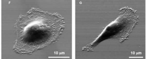
In a test using aggressive metastatic breast cancer cells, they were able to sort the cells based on their mobility. They were then able to collect the cells with high mobility, and run the test again. They found that their mobility was unaffected by repeat tests.
The researchers also found that the more mobile cells had the characteristics and appearance under the microscope of metastatic cells and expressed significantly higher levels of markers associated with metastatic cancer.
While further testing is needed to validate the device, the researchers say it could play an important role in understanding the molecular processes that cause cancer cells to break away from primary tumours and spark metastasis, ultimately informing new therapeutic strategies. It could also benefit research into wound healing, where cells need to quickly congregate at an injured site.