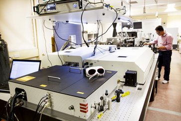
Australian scientists have found a way to image nanoscale structures in high resolution using an extreme UV laser system that fits snugly on a table - a set-up that's far cheaper and more convenient than the multi-million-dollar facilities currently required to image such small objects.
The researchers from Swinburne University of Technology in Australia used the set-up to generate bright beams of coherent extreme UV radiation, that can be exmployed to image structures on the nanoscale.
Conventional optical microscopes aren't able to see structures this small because they're limited by the relatively long wavelengths of light they use to illuminate the sample.
"One way to achieve higher spatial resolution is to use radiation with shorter wavelengths such as extreme UV radiation or 'soft' x-rays," lead researcher Lap van Dao said in a press release.
Currently, the only way to generate intense beams at such short wavelengths is by using large-scale, expensive facilities, such as synchrotrons or free-electron lasers. But the new system developed by the Swinburne researchers offers a much more cost-effective and convenient solution.
The table-top set-up works by producing two beams of high-intensity laser pulses at different wavelengths - one at 800 nm and one at 1,400 nm. Both of these have much longer wavelengths than extreme ultraviolet radiation, which is in the range of 10 to 100 nm, but by illuminating a gas such as argon and overlapping the two beams, the researchers are able to create a bright beam of coherent extreme UV radiation.
This is because the first beam generates 'high-order harmonics' in the extreme UV wavelength region, and then the second beam overlaps to amplify the extreme UV radiation, through a process known as optical parametric amplification, producing a bright, coherent extreme UV beam.
Once this beam has illuminated the structure being imaged, the radiation is scattered slightly, and the resulting 'diffraction' pattern is picked up by a detector on the other side. A computer is then able to reconstruct a high-resolution image of the nanostructure. This is a process known as coherent diffractive imaging, and it eliminates the need for a lens altogether.
The fact that the team has been able to achieve this process using something that can fit on a table is a pretty huge deal, and it suggests that they could do the same to create beams of even shorter wavelengths.
"This research paves the way for the generation of intense radiation at still shorter wavelengths and ultimately to apply coherent diffractive imaging techniques to nano-scale structures and to biological samples in the water window region (2-4 nanometres)," said one of the research team, Peter Hannaford.
The research has been published in Nature Communications.
Love science? Find out more about studying at Swinburne University of Technology.