Electron microscopes have been capable of taking snapshots of individual atoms for nearly half a century. But we've never seen anything quite on this scale.
A new method for catching and measuring the spray of electron beams is giving us a whole new resolution of the sub-ångström world, opening the way to studying molecular structures that would be impossible to see using existing methods.
Last year, engineers at Cornell University in the US performed the equivalent of eye surgery on the traditional electron microscope, ditching the need for corrective lenses and improving the way the eye itself collects and measures light.
Now we have evidence of exactly what that technology can achieve, measuring the bonds between atoms with unprecedented clarity.
At a fundamental level, all microscopes work in a fairly similar way – an object is showered in waves of energy, which are collected and arranged in such a way that we can deduce its shape. Smaller waves mean smaller details.
Electrons can have pretty small wave-like properties that depend on the energy they contain, making them perfect for seeing extra small objects. Instead of lenses, they're focussed using electromagnetic fields.
Aberrations in these fields can limit the size of objects we can see, much as deviations in lenses can blur images. Engineers usually fix these with the electron microscope equivalent of glasses, adding corrective devices to 'fix' the picture.
This fix only goes so far, though. Multiple aberrations demand additional devices, which could theoretically pile up to the point that it's an engineering nightmare.
A device called an electron microscope pixel array detector (EMPAD) does away with the need for these 'glasses' by taking another approach. It's a catcher's mitt for electrons that bounce off the sample made up of a 128 x 128 array of electron-sensitive pixels.
Rather than build an image based on the location of the electrons, it detects the angles of each electron's reflection.
Working backwards using a technique usually applied to X-ray microscopy called ptychography, it's possible to build a four-dimensional map that tells not only where the electrons came from, but their momentum as well.
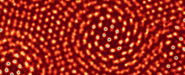
The team put the combination of EMPAD and ptychography to the test by analysing the structure of two stacked sheets of molybdenum disulfide, each a single atom thick.
By rotating one sheet a few degrees, they could compare distances in overlapping atoms, setting a record of resolving a distance of just 0.39 ångströms.
"It's essentially the world's smallest ruler," says physicist Sol Gruner.
The lattice (pictured above) was so clear, they spotted a single missing sulphur atom.
But apart from bragging rights, the technique has another massive advantage.
Electron waves can be made smaller by pumping up their energy. More energy means shorter wavelengths. State-of-the-art microscopes can emit streams of electrons at 300 kiloelectronvolts that can resolve details just under 0.05 nanometres, or 0.5 ångströms.
But more energy can also turn those electrons from a gentle sprinkle of particles into a machine gun burst, putting molecules at risk of disintegrating.
Since this beam was a gentle 80 keV, the electrons weren't energetic enough to break up the structure of the molybdenum disulfide sheets, as they might in a more traditional setup.
Lower energy electron beams mean we can now study bonds in delicate molecules like never before, giving electron microscopy a more gentle touch while providing a whole new level of detail.
This is some artwork we look forward to hanging on our wall.
This research was published in Nature.