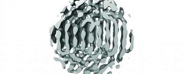
Researchers have developed a new way to capture images of the 3D structures of nanocrystals, tiny particles which hold the promise of fighting cancer and collecting renewable energy.
They are so small their structure is hard to work out and it is difficult to find out how they work. Their dimensions are measured in nanometres, one millionth of a millimetre.
The international research, published in the journal Science, was co-led by Associate Professor Hans Elmlund from the ARC Centre of Excellence in Advanced Molecular Imaging at Monash University.
The imaging method, called 3D Structure Identification of Nanoparticles by Graphene Liquid Cell EM (SINGLE), improves on previous techniques by combining three components.
The first is a graphene cell, a bag one molecule thick which can hold liquid inside it while being exposed to the ultra high vacuum of an electron microscope.
The second is an electron detector which is even more sensitive than traditional camera film and can be used to capture movies of spinning nanoparticles.
Finally, a 3D modelling approach known as PRIME allows using the movies to create three-dimensional computer models of individual nanoparticles.
The next step in the project will include investigating the formation and evolution of nanoparticles and characterising the transitions they go through to reach their final form.
"It is important for us to understand this, so that we can design new materials, for example, to build better or more efficient solar cells, or make better and more economical use of fossil fuels," Elmlund says.

This article was originally published by Business Insider.
More from Business Insider: