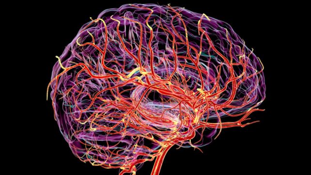
The subtle pulses of tiny blood vessels in the brain can now be mapped in greater detail than ever before using a new imaging method.
It's the first noninvasive tool to measure the expansion and contraction of microscopic arteries and capillaries in the living human brain.
Previous studies suggest that arterial pulses in the cerebrovascular system are predictive of dementia or cognitive decline. This technique could reveal whether that's true or not.
Scientists can now identify dynamic changes across the entire cerebrovascular tree, from the largest branches to the tiniest twigs and offshoots at their ends.
Related: Just One High-Fat Meal Can Disrupt Blood Flow to Your Brain, Study Finds
"Arterial pulsation is like the brain's natural pump, helping to move fluids and clear waste," explains neurologist and senior author Danny Wang from University of Southern California (USC).
"Our new method allows us to see, for the first time in people, how the volumes of those tiny blood vessels change with aging and vascular risk factors. This opens new avenues for studying brain health, dementia, and small vessel disease."
The method was developed at the University of Southern California in collaboration with the medical technology company Siemens Healthcare US. It combines two MRI techniques – vascular space occupancy (VASO) and arterial spin labeling (ASL) – to measure subtle changes in the volume of the brain's vasculature.
The increased pulsation of arteries in the brain is closely linked to cognitive impairment, and some scientists think it might contribute to certain forms of dementia, like Alzheimer's disease. But studies so far have been limited in what they can glean from living human brains, meaning that much research is based on animals.
Wang, lead author and neuroscientist Fanhua Guo, and their team at USC hope their new technique will help fill that gap. They analyzed microvascular pulses in the brains of 11 young participants, in their 20s and early 30s, and 12 older participants in their mid-50s and early 60s.
Ultimately, researchers found pulses of blood in deep white matter sped up with age, and those older individuals with hypertension showed more changes than most.
The findings align with recent animal studies, which suggest that the vasculature of white matter in the brain (composed of nerve fibers) pulses more with age or disease.
"Our method shows strong potential for both research and clinical use, enabling accurate imaging of the cerebral microvascular system and capturing volumetric pulsatility in gray matter and white matter," the authors conclude.
Researchers are not yet sure why this is happening, but they have some hypotheses.
The density of the brain's microvascular system is thought to naturally decline with age, and this reduction in volume and branching may mean that the arteries cannot dissipate the pressures of each pulse as well as they used to.
If that's true, then to release the residual pressure, outer arteries in the brain's white matter may increase the volume of their pulsations.
That in turn could slow the flow of cerebrospinal fluid, which bathes the brain, and which is closely linked to aging and disease.
"These findings provide a missing link between what we see in large vessel imaging and the microvascular damage we observe in aging and Alzheimer's disease," says Guo, who is a postdoctoral researcher in Wang's lab.
The study was published in Nature Cardiovascular Research.