The human brain is a magnificently complex information-processing machine that is extremely off-limits to certain kinds of research.
Science survived the moral void into which brain research fell for much of the 20th century; and, for exceptionally valid ethical reasons, there are now stringent rules governing what researchers are allowed to do with human brains that humans are still very much using.
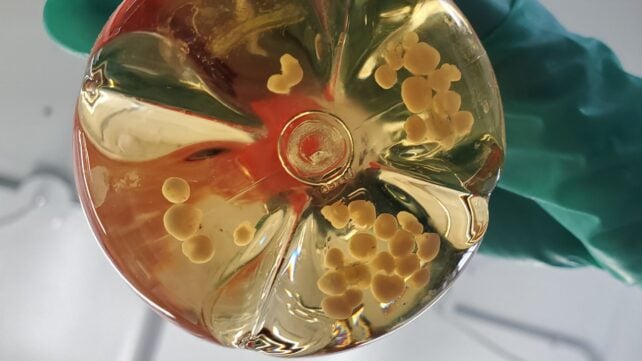
What scientists can do is grow little bits of one in a dish.
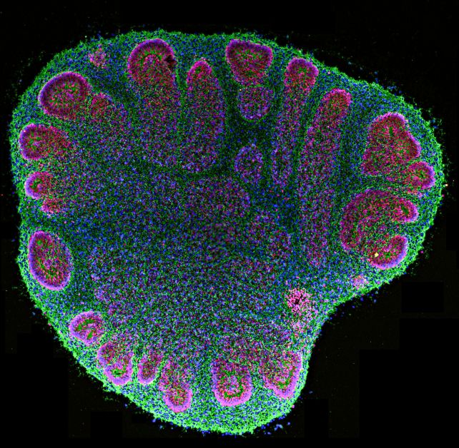
These tiny, three-dimensional clumps of human neural tissue, called brain organoids, aren't whole brains reproduced in a miniaturized form.
But they do reproduce enough of the brain's cellular architecture and electrical activity to give scientists a way to investigate things they could never ethically poke, prod, wire up, and repeatedly zap inside a living person's skull.
Now, a team led by neuroscientist Yoshiho Ikeuchi of the University of Tokyo has grown several such organoids, joined them up in a network, and watched as they 'learned' to distinguish which of their fellows was sending them signals.
"Together, these results demonstrate that repeated input to an organized modular network can rewire organoids generated under identical conditions into functionally differentiated modules," the researchers write, "thereby generating task-relevant heterogeneity that underlies consistent functional enhancement in vitro."

No, the organoids aren't forming sentient brain-blob networks that will rise up and take revenge on their fleshy tormentors.
Rather, the research demonstrates that a particular configuration of modular architecture may be required before neural tissue can learn to tell different signals apart.
The human brain isn't just one homogeneous tangle of neural wiring. It's organized into regions, each with its own local network, which communicate with each other via longer-range connections. This modular organization allows different parts of the brain to perform different roles, while remaining part of a networked whole.
But the minimal requirements for neural tissue to develop functional circuits aren't entirely understood. With the increasing sophistication of brain organoid models, Ikeuchi and his team naturally turned to brain blobs as a means of investigating the problem.
The blobs themselves are grown from human induced pluripotent stem cells (hiPSCs) – basically, mature human cells that have been reprogrammed into a stem-cell-like state, giving them the ability to develop into many different types of cells if coaxed in the right direction.
Ikeuchi and his team coaxed their hiPSCs to develop into brain tissue – giving the researchers a number of tiny chunks of neural tissue to experiment with.
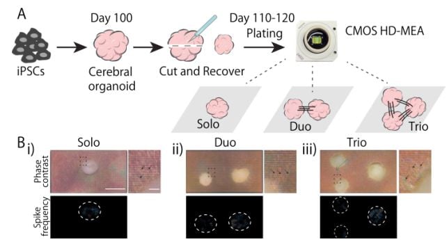
They placed these chunks on special chips covered in tiny electrodes, which allowed the researchers to both stimulate the organoids with electrical signals and record their neural activity.
Some chips got one brain blob. Some got two. And some got three.
The researchers then left them for more than two weeks, giving the organoids grouped on each chip time to grow axons – the long, signal-carrying projections of nerve cells – and form connections.
Recordings taken after this period showed synchronized electrical activity between the organoids that were grouped together, confirming that they had formed functional networks.
Then, it was time for The Zappening (it's a mystery why anyone would think the organoids might want revenge. Complete enigma).
Every day for two weeks, the researchers electrically stimulated the neural networks at two different locations, 100 times at each location, while recording the resulting neural activity elsewhere on the chip.
For the chips with one organoid, the zaps were administered to two places on that single organoid. For chips with two organoids, both sets of zaps went to one, while brain activity was recorded from the other. And for chips with three organoids, the zaps were split between two, while brain activity was recorded from the third, unzapped one.
The idea was to see whether repeated stimulation would eventually teach the neural tissue to respond differently depending on where the signal came from.
To find out, the researchers used machine-learning algorithms to analyze the patterns of brain activity following each zap, and asked them to identify which of the two locations had been stimulated.

At the beginning of the experiment, the researchers deliberately selected stimulation sites that produced responses that were difficult to tell apart. The algorithms could identify the source correctly only around half the time – consistent with what you'd get from guessing.
After The Zappening, a different picture began to emerge.
Not for the single organoids and the pairs – their responses to the two signals remained difficult to tell apart.
But in the three-organoid networks, the two signals became increasingly distinguishable from the activity they produced in the third organoid. The responses also became faster, more stable, and more spatially differentiated.
And three-organoid networks that weren't subjected to The Zappening didn't show the same improvement, suggesting that the change wasn't simply the result of the blobs spending another two weeks growing in a dish.
The repeated signals themselves appeared to have driven the neural networks to reorganize.
It's a very basic form of learning, and a very long way indeed from the complex information processing performed by an actual, fully fledged human brain.
But the results, the researchers say, suggest that organizing neural tissue into connected modules with different roles may help it develop the ability to distinguish between different sources of information.
Since basic signal discrimination is a fundamental requirement for more complex information processing, the result offers researchers a new way to investigate how increasingly sophisticated neural functions emerge.
"Testing organoids with more complex signals and sophisticated tasks will be critical for future research," the researchers write.
"Future studies should focus on identifying potential 'node cells' or 'hub neurons' that facilitate connections within and between organoids – combining this with biochemical and molecular analyses could reveal the mechanistic basis of changes in synaptic plasticity and connectivity."
The research has been published in Communications Biology.
This article was fact-checked by Rebecca Dyer and edited by Rebecca Dyer. While we pride ourselves on our process, we are only human. If you spot a mistake, please let us know.