
A tiny, 2,100-year-old mummy from ancient Egypt had long been thought to contain the remains of a treasured bird - which would make sense, considering the hawk-themed decorations and small size.
But researchers conducting a CT scan have found something else entirely - the remains of a severely malformed human foetus, stillborn at no later than 28 weeks.
The mummy had been in storage at the Maidstone Museum in Kent, England, listed in the inventory as EA 493 Mummified Hawk, Ptolemaic Period.
The funerary casement was the perfect size for a bird, bearing the head of a hawk painted in gilt and hieroglyphics referring to Horus, the falcon-headed sky god of the ancient Egyptians.
In addition, the mummification of animals - from crocodiles to cats to kestrels to scarab beetles - was a very common practice in ancient Egypt. So the mummy did not stand out as anything particularly special or unusual.
It nearly didn't even get CT scanned. The museum was having a human mummy scanned in 2016, and figured it may as well put in a few animal mummies from its collection to be scanned too.
 (Maidstone Museum UK/Nikon Metrology UK)
Arms crossed over the chest revealed that it wasn't a falcon after all - but the scan wasn't very detailed, and the museum experts thought it might have been a monkey.
Then, anthropologist Andrew Nelson of Western University in London, Canada called in a multidisciplinary team to run and analyse an extremely high resolution micro-CT scan.
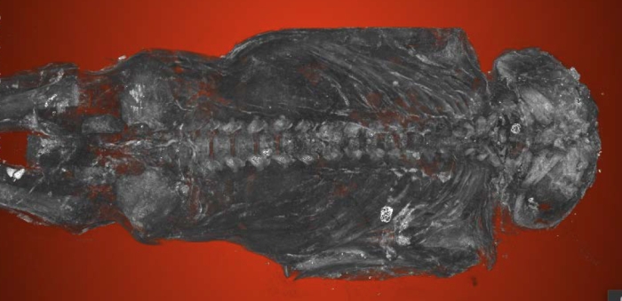
The team discovered the bones belonged to a human male foetus, between 22 and 28 weeks gestation, with severe spinal abnormalities and a rare birth defect that prevents the brain and skull from developing properly.
"On the basis of the highest resolution scan of a foetal mummy ever made, we've been able to determine this individual was severely anencephalic. It would have been a stillbirth, it would not have lived through birth." Nelson said.
"The whole top part of his skull isn't formed. The arches of the vertebrae of his spine haven't closed. His earbones are at the back of his head."
The scans reveal normally formed finger and toe bones, but so serious is the skull deformation, that the brain would have been practically nonexistent. He also had a cleft palate and a cleft lip.

He's also a rarity - one of only eight known mummified foetuses, and only the second every discovered with anencephaly. The first was described in 1826 - nearly 200 years ago. No others have been found since, until EA 493.
The way the remains were preserved mean his family regarded him as special.
"It would have been a tragic moment for the family to lose their infant and to give birth to a very strange-looking fetus, not a normal-looking fetus at all," Nelson said.
"The family's response was to mummify this individual, which was very rare. In ancient Egypt, fetuses tended to be buried in pots, below house floors, in various ways. There are only about six or eight known to have been mummified. So this was a very special individual."
The researchers believe the foetus may hint that the mother's diet was low on foods that provide folic acid, a vitamin that plays an important role in neural tube development. Its intake has been linked with a lower risk of anencephaly.
But it also raises new questions - such as why the mummy was decorated with bird imagery. Other items that may have been buried with the mummy could provide clues, but its provenance is sadly unknown.
Nelson presented the research at the World Congress on Mummy Studies, which was held in Tenerife, Spain on 21-25 May.