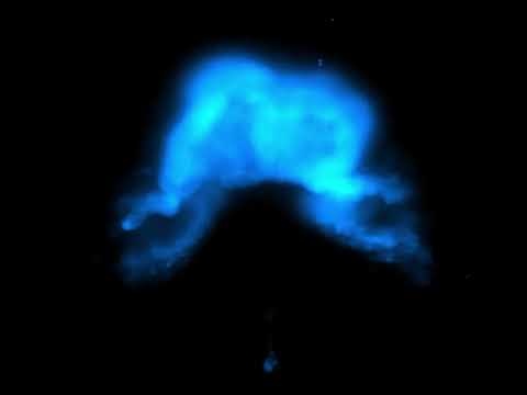
Scientists have caught the intricate dance of cardiac cells coming together to build a heart, in a mesmerizing new timelapse taken during the development of a mouse embryo.
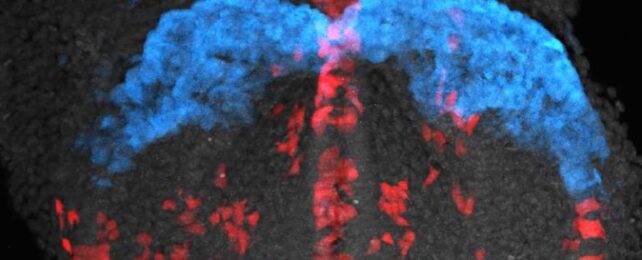
The images were captured using a technique called light-sheet microscopy (LSM). Essentially, LSM involves scanning a sample with a thin sheet of light, creating sharp, detailed, three-dimensional images of living tissue without damaging it.
Researchers from University College London (UCL) and the Francis Crick Institute in the UK used the method to track how mouse embryo cells begin to specialize into roles, divide, and arrange themselves into the structure of a heart.
The team tagged the different types of cells with fluorescent markers, then captured images of them every two minutes for up to 41 hours. The resulting timelapse shows a ragtag group of nondescript cells coming together to form a living, beating mouse heart in a way that's truly captivating to watch.
It's not just beautiful; it helped the team uncover new details about cardiac development. Surprisingly, individual cells seemed to already 'know' where they need to go and which roles they'll end up playing, even as early as four or five hours after the first embryonic cell divided.
"Our findings demonstrate that cardiac fate determination and directional cell movement may be regulated much earlier in the embryo than current models suggest," says Kenzo Ivanovitch, developmental biologist at UCL.
"This fundamentally changes our understanding of cardiac development by showing that what appears to be chaotic cell migration is actually governed by hidden patterns that ensure proper heart formation."
While it's a long way off from any practical benefits, a better understanding of this process could potentially lead to new treatment options for congenital heart defects, the team says.
The research was published in The EMBO Journal.