The biggest barrier to understanding the human brain is the human around it.
Scientists can't exactly tinker around in someone's gray matter while that someone is still using it. Which means many processes involved in brain development and wiring, and what happens to make it go awry, are still poorly understood.
Researchers have spent years finding other ways to study human brain tissue, from cells grown in dishes to tiny three-dimensional structures called organoids. But even the most sophisticated organoids have something missing – the body that a brain normally drives.
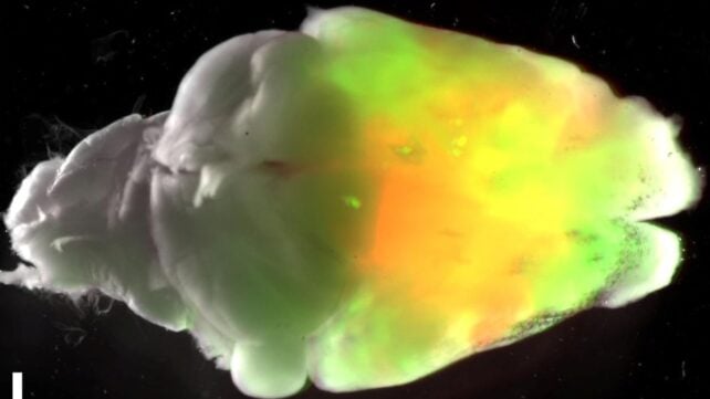
Now, neuroscientists at Stanford University have taken a huge step towards solving that problem. They grew human cortical tissue inside mice whose own cerebral cortex – the outer layer of the brain – had largely been genetically depleted early in development.
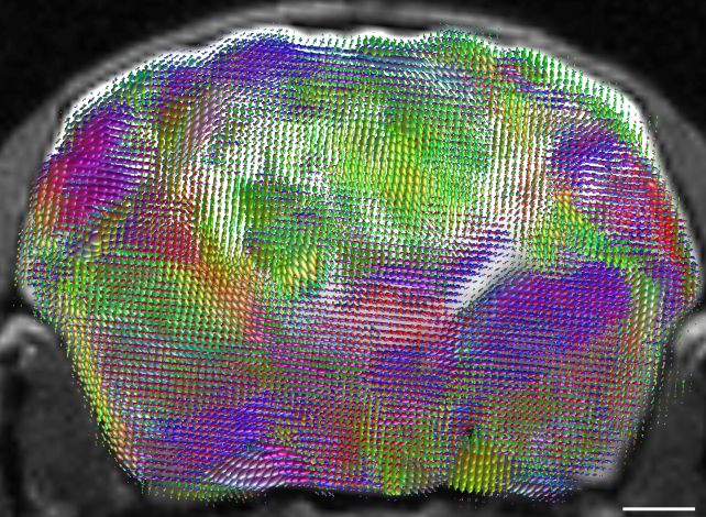
Within three months, the human tissue had expanded to make up more than 90 percent of the cortical tissue by volume. By this point, it contained blood vessels, had wired itself into the mouse nervous system, became electrically active, and sent some projections all the way into the spinal cord.
"The most important point is that these are still mice," neuroscientist Sergiu Pașca of Stanford University told ScienceAlert.
"They have a mouse nervous system, mouse sensory organs, and mouse subcortical structures. What is unusual is that most of the cortical tissue present in these animals is human-derived and that the human neurons grow, integrate, and form functional connections with the rest of the mouse nervous system."
Brain organoids have become very sophisticated in recent years.
Scientists can now grow blobs of brain tissue containing a broad range of the cell types found in the developing human nervous system, and even join organoids representing different regions together into more complex structures called assembloids.
But there's a limit to how far a petri dish can take you. An isolated organoid has no blood supply, no sensory information to process, and no muscles to direct.
This is a problem Pașca and his colleagues have been trying to solve for years. In 2022, the team transplanted human cortical organoids into newborn rats, where the human neurons matured, integrated with the rats' brains, and even responded to sensory information from their whiskers.
But there was still a problem. Human neurons develop far more slowly than rodent neurons. The two developing systems were competing for the same territory. So, the researchers removed the competition.
To do that, the team genetically engineered mice so that most of the cerebral cortex and hippocampus never developed. They called these animals "apallial" mice.
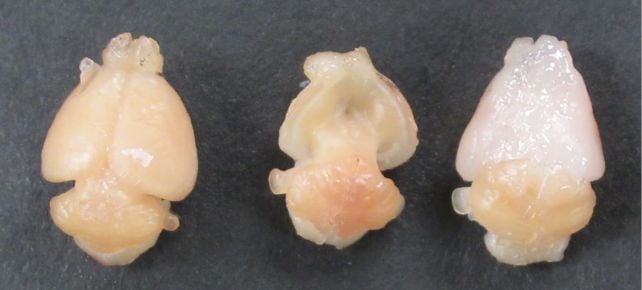
Some were raised that way into adulthood. Others received transplants of human cortical organoids shortly after birth, creating what the researchers call "xenocortical" mice.
As the mice grew and matured over the following months, the grafts grew with them, expanding dramatically through the vacant cortical space.
The resulting tissue wasn't a replica of a human cortex. Even after around six months, it remained developmentally immature, roughly comparable to the human cortex around mid-gestation, without the organized layers of a mature cortex.
Even so, it started to produce neurons that the researchers had struggled to generate in other models.
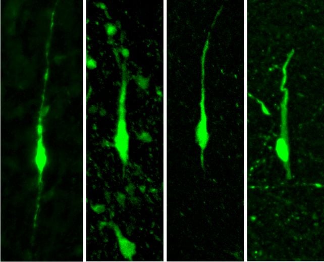
Among them were a group called layer 5 extratelencephalic (L5-ET) neurons, long-range projection neurons that normally connect the cortex with distant parts of the nervous system.
And within that population, the researchers found cells resembling von Economo neurons (VENs) – unusual, elongated neurons found in specific regions of the human brain that have proven particularly difficult to reproduce in the lab – and, notably, were not detected in the earlier rat study.
So what is it about the mouse brain that encourages them to develop?
"We do not yet know, and I think this is one of the most interesting questions raised by the study," Pașca said.
"It is … possible that giving human cortical neurons access to distant targets in other parts of the nervous system provides signals that are difficult to reproduce in an organoid in a dish.
"Another possibility is that there are factors present in the in vivo environment that are missing from organoids in a dish. These could include trophic or growth factors, signals associated with neuronal activity, vascularization, or other interactions with surrounding cells."
But knowing what happens to the human brain tissue was only half of the question. The other half was – what the heck does it do to the mice?
To find out, the researchers put three groups of mice – a normal, unedited control group, the ungrafted apallial mice, and the xenocortical mice – through a Y-shaped maze test.
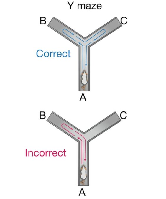
This is because mice naturally prefer to explore a different arm of a maze from the one they just visited – so remembering where they've already been provides a simple test of working memory.
The control mice performed better than chance. The apallial mice didn't, suggesting that without most of their cortex and hippocampus, they had trouble remembering which arm they'd just explored.
The xenocortical mice, however, also performed above chance.
"That is an intriguing result, particularly because the apallial mice did not show the same performance," Pașca said. "But we should be cautious about concluding from this alone that the human neurons are directly responsible for the behavior."
That's a question that will need to be addressed in future research.
For now, the work answers a more fundamental question the scientists were trying to address – not "can human brain cells make mice do better in mazes?" but "can human brain grafts in mice give us a way to study what happens when the brain goes wrong?"
And that could have some very important applications.
"One obvious direction is frontotemporal dementia. Because we can now generate VEN-like neurons in this setting, we can derive cortical organoids from patients, ask whether these rare neurons show selective vulnerability, and test whether candidate interventions can protect them," Pașca explained.
"For epilepsy, we can ask whether disease-associated human neurons generate abnormal circuit activity or seizures and whether those abnormalities can be corrected therapeutically.
"For autism and other neurodevelopmental disorders, we can ask whether defined alterations in human cortical circuits lead to reproducible functional or behavioral consequences in the animal."
But integrating human neural tissue into another animal on this scale inevitably raises some difficult ethical questions.
The team didn't wait until the experiments were finished to start asking them. In addition to the usual animal research review, Pașca said Stanford bioethicists and an external committee of neuroscientists, ethicists, legal scholars, and patient advocates were involved.
One question is about the animals themselves – whether their suffering is justified by the type of research performed, or whether the information can be obtained another way.

"Another important question is whether introducing increasingly complex human neural tissue into an animal nervous system could lead to unexpected, emergent, or novel properties that would require additional ethical consideration," Pașca said.
"We considered this possibility explicitly and monitored the animals carefully, both biologically and behaviorally, as the work progressed."
Related: Scientists Grew Stem Cell 'Mini Brains' And Then The Brains Sort of Developed Eyes
At the same time, there is an ethical cost to not developing better models for neurological and psychiatric disease.
"Neurological and psychiatric disorders affect nearly one in five people, and for many of these conditions we still do not understand the underlying biology well enough to develop effective treatments," Pașca said.
"Many of these disorders involve human neural circuits and behavioral consequences that cannot be adequately modeled in conventional animal models or in isolated human cells."
The findings have been published in Nature.
This article was fact-checked by Rebecca Dyer and edited by Rebecca Dyer. While we pride ourselves on our process, we are only human. If you spot a mistake, please let us know.