In a move ripped straight from the pages of a science-fiction novel, physicists in Germany have developed an optical tractor beam that can trap biological cells, such as blood, algae, and bacteria, inside a laser beam for high-resolution study.
Before now, scientists had to mount these cells on substrates (think: glass slides) to observe them under a microscope, causing them to be damaged and altered before they could be studied. The new 'tractor beam' system negates all of that, allowing for more accurate observations of living cells in their natural state.
"Our new method enables us to take cells that cannot be anchored on surfaces and then use an optical trap to study them at a very high resolution. The cells are held in place by a kind of optical tractor beam," said team leader Thomas Huser, from Bielefeld University in Germany.
"The principle underlying this laser beam is similar to the concept to be found in the television series Star Trek."
The system works by using infrared lasers to hold on to cells in a fluid solution, allowing them to float like they would in nature. Normally, cells would be mounted onto slides - a process that can damage or kill the cell, leading to incomplete results
"The laser beam is very intensive but invisible to the naked eye because it uses infrared light," said team member Robin Diekmann.
"When this laser beam is directed towards a cell, forces develop within the cell that hold it within the focus of the beam."
The whole system works by trapping these cells inside the beams of laser light, using two separate lasers to manipulate them, allowing the team to turn them over or around to capture a complete view on all sides.
"What's special is that the samples are not only immobilised without a substrate but can also be turned and rotated. The laser beam functions as an extended hand for making microscopically small adjustments," Huser explains.
Taking this method a step further, the team also developed the system for use in super-resolution fluorescence microscopy - a method that involves adding fluorescent probes to the specific cells, and lighting them up with a zap from a laser beam.
With the cell illuminated, delicate sensors affixed to the microscope can measure the amount of radiation coming from within the cells, allowing them to map the entire structure in 3D.
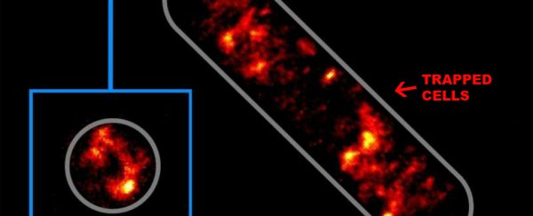
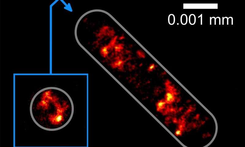
So far, the team has used their new technology to hold in place bacterial cells, rotating them around to measure all the sides.
They were able to observe the structure of the bacteria's DNA in 3D at a resolution of 0.0001 millimetres. To put that into perspective, a hair is about 0.06 millimetres wide.
 Bielefeld University
It's hoped that the new tractor beam will help researchers to better understand how cells operate when studied in more natural conditions - an environment that can't be replicated using glass slides.
The next step for the team is to figure out a way to observe interactions between living cells, which has the potential to shine a new light on how cells work inside the body. If they are successful, we might be on a cusp of learning a whole bunch of new biological information.
The team's work was published in Nature Communications.