
Imagine if scientists could look inside the entire human body at once, into all the tissues and organs, and watch as medicine winds its way through every inch of our being.
Think of the possibilities: such capacity could allow us to diagnose and track disease like never before, not to mention the advantages for drug development.
It may sound like a pipe dream, but after 13 years of work, a team led by researchers at the University of California Davis has designed the first machine with the potential to do just that.
Combining positron emission tomography (PET) and X-ray computed tomography (CT), the new machine can capture three-dimensional images in as little as one second, and in greater detail than ever before.
The invention could have a profound impact on medicine, allowing scientists to see the outlines of cancers and other diseases far more easily, as well as track their movement throughout the body.

"While I had imagined what the images would look like for years, nothing prepared me for the incredible detail we could see on that first scan," said one of the researchers, biomedical engineer Simon Cherry.
"While there is still a lot of careful analysis to do, I think we already know that Explorer is delivering roughly what we had promised."
That's a big statement, because Cherry and his colleague, radiologist Ramsey Badawi, have promised quite a lot. Three years ago, after receiving a US$15.5 million grant from the National Institutes of Health (NIH), Badawi compared the possibilities of Explorer to the Hubble Space Telescope.
Except, instead of letting us behold far off galaxies, he claimed this invention would let us peer into the infinite complexity of the human body.
"In many ways it wasn't known what the Hubble would see before it went up there, and I feel the same is true with explorer," said Badawi at the time.
"It's vastly exciting because we've never been able to look at the body in this way, so we don't actually know what we're going to end up seeing and that could cause revolutions."
It's certainly a bold claim, but the initial results look extremely promising. Even Badawi, who had high hopes from the start, said he was dumbfounded by Explorer's first images.
"The level of detail was astonishing, especially once we got the reconstruction method a bit more optimised," he said.
"We could see features that you just don't see on regular PET scans."
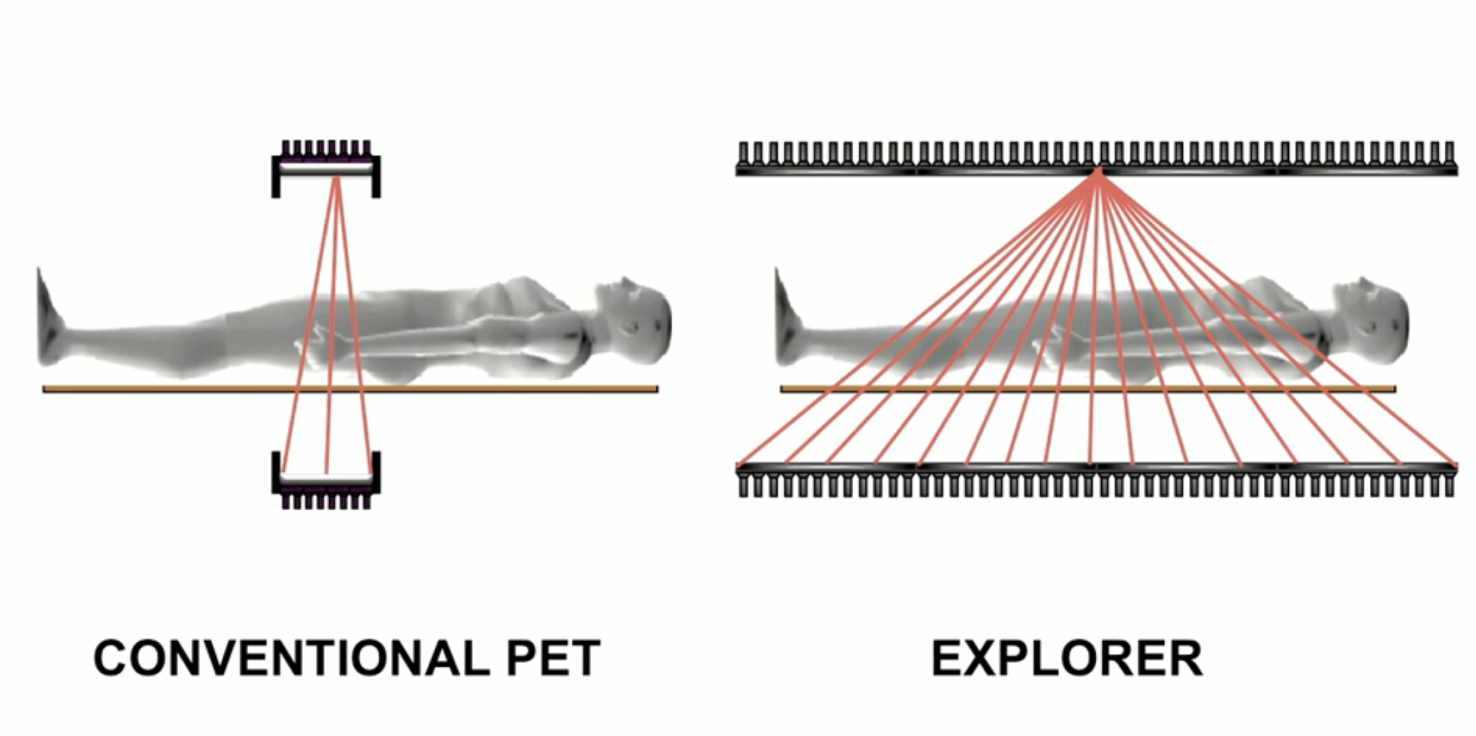
The new machine is so efficient, it can scan the whole body in as little as 20 to 30 seconds, up to 40 times faster than current PET scans. The instantaneous snapshot will allow scientists to track specially-marked drugs as they travel through the human body over time, a result the researchers described as "mind-blowing."
"There is no other device that can obtain data like this in humans, so this is truly novel," said Badawi.
Even better, the new scanner is safer than current PET scans, delivering a dose of radiation that is 40 times less.
When using current scanning technology, scientists have to worry about controlling cumulative doses of radiation, as they can be harmful to human health. Explorer could now make it feasible for individuals to undergo multiple scans and improve its use for children, who are particularly vulnerable to radiation.
"… in all cases, we can scan better, faster or with less radiation dose, or some combination of these," Cherry said.

This sort of machine could prove to be useful in just about any field of medicine. The ability to examine organs and tissues in the body at the same time will allow us to measure blood flow more precisely than ever before. We could literally watch every facet of our body take up glucose.
The researchers are hoping that the scanner will improve the study of cancer and a whole variety of other diseases causing inflammation, infection, immunological or metabolic disorders in the body.
They say the machine could be ready for use by as early as June of 2019.
"I don't think it will be long before we see at a number of Explorer systems around the world," said Cherry.
"But that depends on demonstrating the benefits of the system, both clinically and for research. Now, our focus turns to planning the studies that will demonstrate how Explorer will benefit our patients and contribute to our knowledge of the whole human body in health and disease."
The first images will be shown at the upcoming Radiological Society of North America meeting starting on 24 November 2018.