When you move to a new place, one of the first things you might do is build yourself a cozy nest – a space of your own where everything is just the way you want it to be.
Cancer, as it turns out, can do something eerily similar.
As a tumor grows, it alters its surrounding environment to suit its own needs – pushing tissue aside, remodeling blood vessels, and hijacking the body's functions to support its continued existence and growth.
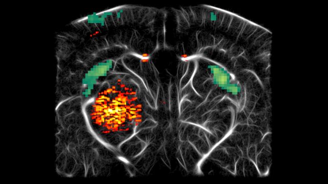
Now, a team of scientists has developed a new ultrasound technique that allows them to watch this nesting process in action as a deadly tumor lodges itself deep in the brain of a mouse.
The tumor is glioblastoma, and the way it builds its nest may offer clues about how to fight it.
"The ability of our platform to capture all three dimensions of tumor progression in vivo in awake animals represents a major step forward in studying neuro-oncological processes with spatiotemporal precision and developing improved treatments," writes a team led by chemical engineer Mikhail Shapiro of Caltech in a paper published in iScience.
There's no good kind of cancer, but some are distinctly nastier than others. One of the nastiest is a group called gliomas – typically malignant growths that arise from glial cells or their precursors in the brain or spinal cord.
The worst form is glioblastoma, which has a 5-year survival rate of just 5 to 7 percent.
Part of the reason gliomas are so difficult to treat is that they commandeer critical brain functions for their own ends, inextricably tangling with surrounding healthy tissue and blood vessels to fuel their own survival.
It's also difficult to understand how the process unfolds. The brain is a highly complex and organized organ. Scientists can't just go poking about to see how it works; even in animal models, the poking can cause damage that changes the very thing they're trying to observe.
What we do have is non-invasive imaging technology that can help peer inside the living brain, but even then, there are limitations.
Magnetic resonance imaging (MRI), for example, can image the entire brain and track both tumors and blood vessels, but isn't well suited to studying brain function in awake, behaving animals. Optical imaging offers exquisite detail, but can't penetrate very deeply into the brain.
And no single technique can show scientists everything they need to see at once.
"Critically," the researchers write, "no existing modality captures tumor growth, vascular remodeling, and the hemodynamic correlates of neural activity together within a single, co-registered acquisition at comparable spatial resolution and depth."
So, they decided to try to combine three techniques into one ultrasound technique that captures more of the evolution of glioblastoma than previous attempts have yielded.
The first hurdle to clear was how to make the glioblastoma stand out among the surrounding brain tissue in the ultrasound images.
The solution was a particularly clever bit of biological engineering. Some bacteria naturally produce little bubbles of gas called gas vesicles. The researchers grew human glioblastoma cells and engineered them with the bacterial genes that perform this effervescent trick.
Then, they embedded the glioblastoma cells in a hydrogel phantom – basically, a medium with a tissue-like consistency that allowed them to test whether the bubbles helped the glioblastoma material stand out under ultrasound.
Tick that box as accomplished. Now all they had to do was pop it in a brain. Igor, fetch the mice.
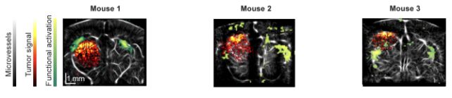
To test the system in living brains, the researchers implanted their engineered human glioblastoma cells in the left thalamus of three mice.
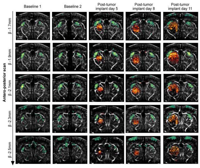
Then, over a period of 11 days, the team repeatedly used trimodal ultrasound to observe the tumors as they took root and grew.
One mode used the gas vesicles to reveal the tumor itself. Another tracked changes in blood volume associated with brain activity, while the third used tiny bubbles injected into the bloodstream to map the network of blood vessels around the tumor in extraordinary detail.
And, magnificently, the technique appeared to work.
The mice underwent ultrasound scans while awake and able to move around on a cushioned platform, with their heads held in position to keep the wiggling to a minimum.
As each tumor grew and expanded, the researchers were able to track how it literally shoved the nearby lateral geniculate nucleus (LGN), which processes visual information, out of position.
At the same time, the surge of blood that normally accompanies activity in the LGN when the mouse sees something was diminished on that side, although the drop was only statistically significant in one mouse; in another mouse, it occurred on both sides.
Then, the tumor rearranged the plumbing. Vascular imaging, captured under anesthesia, revealed blood vessels being displaced and reorganized around the growing tumor.
In the one mouse with a complete dataset, blood also moved more slowly through the vessels around the tumor, with signs that some were narrowing or becoming obstructed.
And the tumor seemed to develop its own little microcosm, tucked inside the brain.
As blood volume ebbed and flowed through the surrounding brain in coordinated patterns, the tumor kept its own rhythm, apparently unrelated to what was happening around it.
But at this point, it's not really possible to say exactly what it was doing or why.
The point of this research was to test whether the technique was able to track glioblastoma changes in living, awake mice in granular detail.

Figuring out what those changes mean and whether they remain consistent across a broader number of subjects is going to require further research.
There are some important caveats. This was a proof-of-concept experiment involving just three mice, using a glioblastoma model that doesn't reproduce all the messy complexity of the cancer as it occurs in humans.
The next step will be to test the technique on larger numbers of animals and more realistic brain cancer models to see whether the strange patterns the researchers observed hold up.
In future work, the technique could also be used to watch what happens when treatment is introduced – not just whether a tumor shrinks, but how the brain and blood vessels tangled up with it respond.
Eventually, the researchers hope that watching glioblastoma build its nest will show them how to tear it down – twig by vicious twig.
The research has been published in iScience.
This article was fact-checked by Rebecca Dyer and edited by Rebecca Dyer. While we pride ourselves on our process, we are only human. If you spot a mistake, please let us know.