Did you know you have tiny tunnels in your head? That's OK, no one else did either until recently! But that's exactly what a team of medical researchers have just found in mice and humans - tiny channels that connect skull bone marrow to the lining of the brain.
The research shows they may provide a direct route for immune cells to rush from the marrow into the brain in the event of damage.
Previously, scientists had thought immune cells were transported via the bloodstream from other parts of the body to deal with brain inflammation following a stroke, injury, or brain disorder.
This new discovery suggests these cells have had a shortcut all along.
The tiny tunnels were uncovered when a team of researchers set out to learn whether immune cells delivered to the brain following a stroke or meningitis originated from the skull, or the larger of the two bones in the shin - the tibia.
The specific immune cells they followed were neutrophils, the "first responders" of the immune squad. When something goes awry, these are among the first cells the body sends to the site to help mitigate whatever is causing the inflammation.
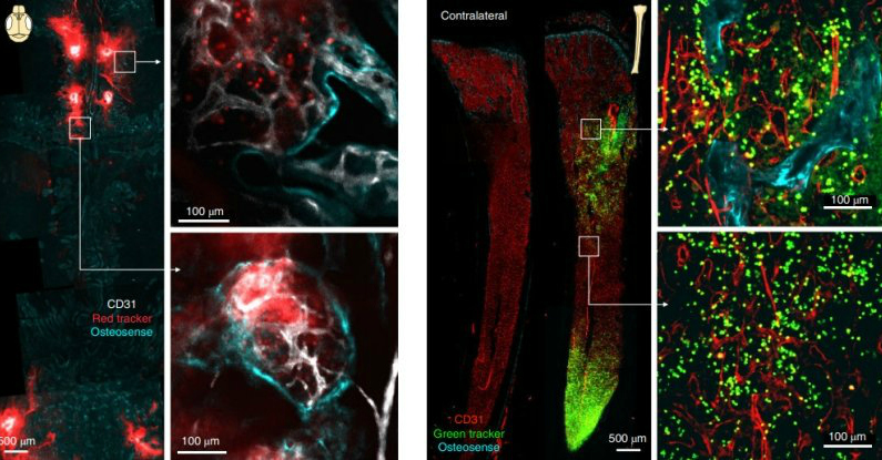
The team developed a technique to tag cells with fluorescent membrane dyes that act as cell trackers. They treated these cells with the dyes, and injected them into bone marrow sites in mice. Red-tagged cells were injected into the skull, and green-tagged cells into the tibia.
 (Herrison et al., Nature Neuroscience, 2018)
Once the cells had settled in, the researchers induced several models of acute inflammation, including stroke and chemically induced meningoencephalitis.
They found that the skull contributed significantly more neutrophils to the brain in the event of stroke and meningitis than the tibia. But that raised a new question - how were the neutrophils being delivered?
"We started examining the skull very carefully, looking at it from all angles, trying to figure out how neutrophils are getting to the brain," said Matthias Nahrendorf of Harvard Medical School and Massachusetts General Hospital in Boston.
"Unexpectedly, we discovered tiny channels that connected the marrow directly with the outer lining of the brain."
 (Herrison et al., Nature Neuroscience, 2018)
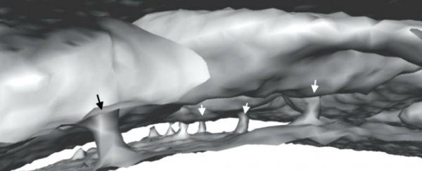
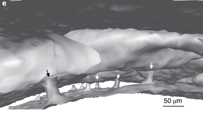
Using organ-bath microscopy - which uses a bath to maintain the integrity of the tissue while it is being examined - the team imaged the inner surface of a mouse's skull. There, they found microscopic vascular channels directly connecting the skull marrow with the dura, the protective membrane that encases the brain.
Normally, red blood cells flow through these channels from the interior of the skull to the bone marrow; but, in the case of stroke, they were mobilised to transport neutrophils in the opposite direction, from the marrow to the brain.
This was in mice, though. To find out if humans have something similar, they obtained pieces of human skull from surgery and conducted detailed imaging.
They noticed channels there as well; five times larger in diameter than the channels in the mouse skulls, in both the inner and outer layers of bone.
It's an amazing discovery, because inflammation plays a role in many brain disorders, and this could help scientists understand more about the mechanisms at play. It could also help understand conditions such as multiple sclerosis, wherein the immune system attacks the brain.
However, further research will need to be conducted to determine the types of cells aside from neutrophils that use these tiny tunnels, and the role they play in various conditions.
The team's research has been published in the journal Nature Neuroscience.