Heart surgery might be one of the most worrying medical procedures patients ever have to undergo, but in the future we could feel considerably safer in the knowledge that surgeons have already planned the operation beforehand – using a highly realistic, personalised 3D model of the blood-pumping organ in question.
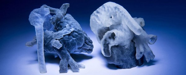
Thanks to a new system devised by researchers at the Massachusetts Institute of Technology (MIT), surgeons are now able to physically assess patients' hearts before opening them up via 3D-printed models that have been sourced from MRI scans. The system produces a tangible model in just a few hours, enabling physicians to prepare for the anatomical idiosyncrasies of individual patients prior to surgery commencing.
"Our collaborators are convinced that this will make a difference," said Polina Golland, a professor of electrical engineering and computer science at MIT, in a press release. "The phrase I heard is that 'surgeons see with their hands,' that the perception is in the touch."
The technique, co-developed with researchers at the Boston Children's Hospital, required several different innovations to bring it to fruition, including software capable of independently analysing MRI scans, together with new procedures to enhance the precision of MRI scans tenfold.
Previous attempts at producing printable models required extensive manual involvement from human experts to determine the boundaries of individual MRI cross sections – hundreds of which can make up a full scan – blowing out the time it took to consolidate the data.
"They want to bring the kids in for scanning and spend probably a day or two doing planning [how] exactly they're going to operate," said Golland. "If it takes another day just to process the images, it becomes unwieldy."
By contrast, the new method produces its strongest results when human intervention is kept to a minimum, with algorithms left to infer the rest. "I think that if somebody told me that I could segment the whole heart from eight slices out of 200, I would not have believed them," said Golland. "It was a surprise to us."
The resulting system, to be presented at the International Conference on Medical Image Computing and Computer Assisted Intervention in Munich, Germany, next month, will now be evaluated by seven cardiac surgeons at the Boston Children's Hospital using 3D models based on the hearts of 10 patients who have already received treatment.
The end goal is to determine whether the 3D models can improve surgical outcomes by contrasting surgical plans based on physical models to operations that were conducted in real-time without the benefit of access to the 3D mock-ups beforehand.
"Absolutely, a 3D model would indeed help," said Sitaram Emani, a cardiac surgeon at Boston Children's Hospital, who was not involved with the research. "We have used this type of model in a few patients, and in fact performed 'virtual surgery' on the heart to simulate real conditions. Doing this really helped with the real surgery in terms of reducing the amount of time spent examining the heart and performing the repair."
Outside of clinical outcomes, another benefit to using personalised physical models is helping show what the patient's heart looks like when explaining medical procedures to patients – and particularly their families when it comes to operations on children.
"[H]aving this immensely simplifies discussions with families, who find the anatomy confusing," Emani said. "This gives them a better visual, and many patients and families have commented on how this empowers them to understand their condition better."