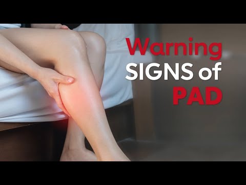
Imagine having blocked arteries in your legs and not knowing it. At first, there may be no symptoms at all. Just occasional fatigue, cramping or discomfort – symptoms easy to dismiss as ageing or being out of shape.
But as blood flow worsens, a small cut on your foot might not heal. It can turn into an ulcer. In the worst cases, it can lead to amputation. This condition is called peripheral artery disease (PAD) – and it's far more common than many realise.
PAD affects around one in five people over the age of 60 in the UK, and is especially prevalent in people with diabetes, high blood pressure, or kidney disease.
PAD is rarely an isolated issue: it's usually a sign of widespread atherosclerosis, the build-up of fatty deposits that can also narrow arteries in the heart and brain.
Related: Your Smartphone Could Soon Measure Your Blood Pressure With Just a Touch
It also significantly increases the risk of heart attacks, strokes and other conditions linked to poor blood flow to vital organs. Research shows that a large proportion of people diagnosed with PAD will die within five to ten years, most often due to these complications.
Early detection is key to reducing the impact of PAD, and I've been working with colleagues to develop a faster, simpler way to diagnose it.
PAD testing
Doctors can check circulation in the feet by comparing blood pressure in the toe with that in the arm. The result is known as the toe–brachial index (TBI). The trouble is that the test needs a toe-sized cuff, an optical sensor and a doctor who knows how to use the equipment.
Many GP surgeries and foot clinics don't have this kit. And in many people, especially those with diabetes or stiff arteries, the test doesn't always give a clear or reliable, result.
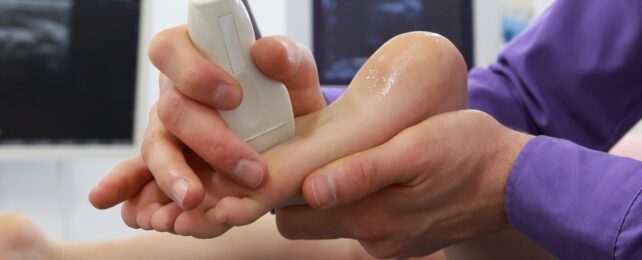
Our research team asked a simple question: could we turn a routine ultrasound scan into a quick, reliable way to measure blood flow in the foot?
Most hospitals, and many community clinics, already have handheld ultrasound probes, which use Doppler sound to track how blood flows through vessels.
This works through the Doppler effect: as blood moves, it changes the pitch of the sound waves. Healthy blood flow creates a strong, steady "swoosh", while a narrowed or blocked artery produces a faint or disrupted sound.
Doctors are trained to hear the difference and use these sound patterns to spot circulation problems, especially in conditions like PAD.
But my research team wondered whether a computer could do more than listen: we wanted to know whether it could convert the shape of that Doppler "wave" into a number that mirrors the TBI.
To investigate, we scanned the feet of patients already being treated for PAD – 150 feet in all. For each artery, we used Doppler ultrasound to measure how quickly blood surged with each heartbeat, a pattern known as the acceleration index. We then compared these results to the standard toe–brachial index, the traditional test that measures blood pressure in the toe.
A one-minute scan, a nearly perfect match
The acceleration index alone was able to predict the standard toe–brachial index with 88% accuracy. Using a simple formula, we converted that Doppler reading into an "estimated TBI" – a number that closely mirrored the conventional result. It needed no toe cuff, no optical sensor and it took under a minute to perform.
Even more encouraging, estimated TBI rose in tandem with traditional TBI results after treatment. When patients underwent angioplasty – a procedure to reopen blocked arteries – their estimated TBI increased almost identically to the measured TBI. That means this scan doesn't just help diagnose PAD; it could also be used to track recovery over time.
Crucially, our approach works with equipment that's already widely available. We repeated the experiment using a basic pocket Doppler: the kind many GPs and podiatrists have tucked in a drawer.
While it wasn't quite as precise as hospital-grade ultrasound, the results were still strong. With some additional software refinement, doctors could soon assess foot circulation quickly and accurately using tools they already own, without adding time to a busy clinic schedule.
Why early detection matters
Because early diagnosis of PAD changes everything. It can mean the difference between losing a foot, keeping your mobility and living longer with a better quality of life. It can shorten hospital stays and reduce the risk of heart attack and stroke.
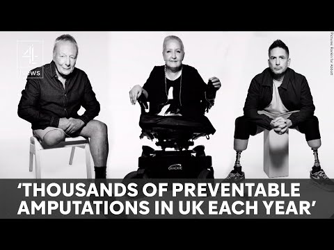
But right now, too many people with PAD aren't diagnosed until they already have chronic limb-threatening ischaemia – the most severe form of the disease. This condition occurs when blood flow to the legs or feet becomes critically low, depriving tissues of oxygen.
It can cause constant foot pain (especially at night), wounds that won't heal and, in advanced cases, tissue death (gangrene) and the risk of amputation. Without urgent treatment to restore circulation, chronic limb-threatening ischaemia can be life-threatening.
Part of the problem is that the tools used to diagnose PAD are often slow, expensive or too complicated for routine use. That's why a simple, cuff-free Doppler scan that provides a reliable estimate of toe–brachial index is so promising.
It uses equipment that many clinics already have, takes less than a minute and delivers immediate results – offering a faster, easier way to spot poor circulation before serious damage is done.
We're now looking at ways to automate the measurement so that it can be used even by non-specialists. We're testing it in various clinics with different patient groups and exploring its performance over time.
But the evidence so far suggests that this could become a key part of vascular care – not just in hospitals, but in GP surgeries, diabetes clinics and anywhere else early intervention could save a limb.
Blocked arteries don't need to stay hidden. With the right tools, we can find them earlier, treat them faster and protect people from the devastating consequences of late diagnosis.![]()
Christian Heiss, Professor of Cardiovascular Medicine, Head of Department of Clinical and Experimental Medicine, University of Surrey
This article is republished from The Conversation under a Creative Commons license. Read the original article.