
What else might be hidden in the electrical language of the cells in our brains?
Neuroscientists often record the activity of neurons as a series of simple events: A cell produces an electrical pulse, or it does not. When and how often those pulses appear can tell researchers a great deal about the brain.
But a new preprint study may have found a detail this approach overlooks.
The millisecond-long electrical sparks produced by neurons are not all identical. Their shapes seem to offer clues about what the cell has recently experienced.
That means an electrical signal may carry more information than the simple binary mark used to represent it.
University of California, San Diego neuroscientist Bradley Voytek told ScienceAlert that these electrical pulses, known as action potentials, look like simple, uniform spikes only when viewed from a distance.
The study has yet to be peer-reviewed, and the researchers urge some caution until it has been. But it adds to a growing picture of how much detail a single neural signal might contain.
"An action potential only looks like a 'spike' when you are zoomed out, but when you zoom in on it, and look at its full waveform, which is only about a millisecond long, the shape of that voltage trace tells you something about what's happened recently to that neuron," Voytek said.
"When we turn an action potential into a binary event, we throw away information about its recent input."
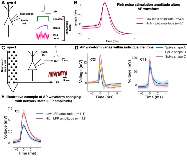
The researchers first examined recordings from two mouse neurons in the part of the brain involved in vision. When the cells received different electrical currents, features changed, such as the height of their signals and how quickly the voltage fell.
Those currents included a steady input, one that gradually increased, and a fluctuating input known as pink noise. Recordings made inside the cells by other researchers captured small changes in each electrical pulse.
In one neuron, a machine-learning model distinguished among three types of electrical stimulation using only the shape of the signal, with 78.4 percent accuracy. There were not enough recordings from the second neuron to run the same test.
The signal's shape also offered clues about how strong or variable the incoming current had been. Some of those clues reached back as far as half a second before the electrical pulse. That is longer than the pulse itself, which lasts about a millisecond.
The researchers then examined brain recordings from rats. This time, they looked at neurons active on their own, rather than cells given a specific electrical current.
In 30 of the 40 neurons examined, signals from the same cell fell into more than one distinct group of shapes.
In a small number of neurons, the differences were so large that signals from the same cell could look as different as signals from separate neurons. In most cells, the differences were smaller.
The researchers also compared signal shape with electrical activity around the cell. In most of the neurons, shape predicted the strength of a nearby electrical signal better than the time between pulses alone.
The relationship was statistically detectable in 87 to 90 percent of cells, although its predictive power was usually modest.
In the cell with the strongest relationship, the shape of its pulses accounted for about 64 percent of the variation in one measure of nearby electrical activity. That result came from a selected cell; most cells showed much weaker relationships.
Does that mean neurons change the shape of their signals to send one another a hidden message?
That is the part of the puzzle still missing.
"Our data can't directly address whether the spike shape is communicating information to other neurons, unfortunately," Voytek said.
What the study does show is that signal shape is linked to the electrical input a cell has recently received and to activity around it.
He points to earlier work showing that changing the width of an electrical pulse can alter how much calcium enters a neuron, which can affect what it passes on to other cells. Whether that happens here still needs to be tested.
The recordings from experiments using different electrical currents came from just two cells. Both datasets were originally collected for other purposes. So we do not yet know how widespread these patterns are in other parts of the brain or among other neuron types.
There is another side to the findings.
Scientists can use the shapes of signals in brain recordings to work out how many different neurons they are listening to. If a single cell can produce signals with different shapes, that task may sometimes be harder than we thought.
The central question remains open. By recording when a neuron produces a signal, we may be overlooking information in the signal's shape. Perhaps another clue to the brain's electrical language lies in the shape of the spark.
A preprint of the study is available on bioRxiv.
This article was fact-checked by Rebecca Dyer and edited by Rebecca Dyer. While we pride ourselves on our process, we are only human. If you spot a mistake, please let us know.