Three years ago, a family in Boston was thrown into chaos during the small hours of the morning. A man, who moments ago had been sleeping soundly next to his wife, was on the floor convulsing, and nobody knew why.
He was confused, uttering nonsensical words and tried to resist being taken by ambulance to Massachusetts General Hospital. There, through a painstaking diagnostic process, doctors discovered an unwelcome brain guest.
On examination, the unfortunate man's heartbeat and breathing were slightly elevated, but toxicology and chest X-rays showed no abnormalities. There was no physical evidence to suggest an underlying chronic disease, no history of illness or unusual behavior in the lead up to the event, nor any known family history of neurological problems.
"The patient also had blood in his mouth, presumably from biting his tongue," writes physician Andrew Cole in a newly published case report.
The 38-year-old man was treated with lorazepam for the seizures, but it took yet more work to find out what had caused them in the first place.
A number of conditions can lead to seizures, or symptoms that look like them. Anything that disrupts blood flow to the brain can cause them, so checking out the entire circulatory system is important. Tests revealed the man's liver, which regulates the chemicals in our bloodstream, and kidneys, which remove waste from the blood and regulate blood pressure, were both functioning properly.
Momentary loss of blood to parts of the brain (ischemic attacks), drugs, migraines, and psychiatric events can also produce seizure-like symptoms, but the toxicology tests and the fact the man had been in perfect health previously, ruled these out.
"Obtaining the clinical history is key," explains Cole. "The most powerful tool in the evaluation of a possible seizure is additional information."
The patient's history did provide a clue. He had migrated from a rural area of Guatemala 20 years ago.
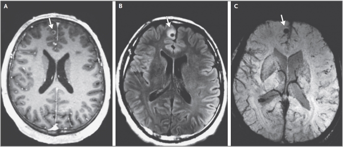 MRI scans focusing on one of the three brain lesions (arrow). (Cole et al, NEJM, 2021)
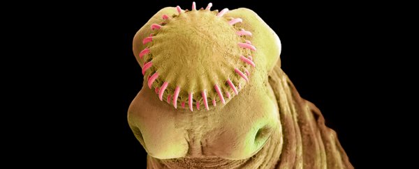
Brain scans revealed three calcified lesions. Given their presentation and the patient's history, the doctors concluded they were cysts belonging to the parasitic pork tapeworm (Taenia solium). These white, ribbon-like worms rely on human hosts to reach the adult phase of their lifecycle, where they latch onto the small intestine with dozens of little hooks
Here, the flatworms feast on surrounding nutrients and can grow up to a shocking eight meters long. This is the stage they're able to sexually reproduce if they get lucky – otherwise they'll reproduce asexually. Their eggs hitch a ride on our feces out to the rest of the world. They can then survive in the environment for up to two months as eggs, in the hope of being eaten by another animal.
Pigs are the most common intermediate host, but sometimes other humans or even the originally infected human eat the tapeworm eggs, too. They hatch in the intestines of their devourer and the resulting larva bore their way into the bloodstream, preferably to nest in tasty pig muscles that can then transport them back to humans who've eaten undercooked meat.
However, the resulting cysts the larva form can develop in any organ, and it is these that cause the most severe problems – especially if they establish themselves in the brain.
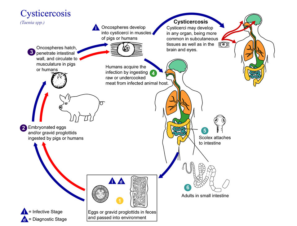 (Alexander J. da Silva/Melanie Moser/CDC)
This condition is called neurocysticercosis and it's the leading cause of acquired epilepsy in many parts of the world – including Latin America and sub-Saharan Africa. Thousands of people in the US alone are thought to present cases not unlike this one each year.
Neurocysticercosis is a preventable disease, but despite its prevalence and severity, relatively little resources have been devoted to combating it, leading the CDC to class it as a neglected tropical disease.
Preventatives include thorough hand washing, cooking and handling meat safely, and prompt treatment for people with intestinal tapeworms.
Some cases of neurocysticercosis require surgery to remove a problematic cyst from the brain, as in a recent case of a 25-year-old Australian woman, who experienced persistent headaches and blurred vision. As the cysts can form in different parts of the brain, symptoms can vary greatly.
As for this case, the man was treated with anti-inflammatory, anti-seizure, and two antiparasitic medications. He was released from the hospital with no remaining symptoms after five days, and remains seizure free three years later.
However, he'll likely need to continue taking the anti-seizure medication.
"The issue of when to stop the medication is problematic, because the calcified lesion will remain in perpetuity," says Cole.
This case study was reported in The New England Journal of Medicine.