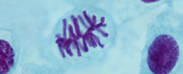
If you've ever studied any chemistry or biology, there's a very good chance you've come across the common pictorial representation of what a chromosome is supposed to look like.
As millions of high-schoolers and undergraduates will attest, it's a tall, narrow X-shape - visualising what two joined chromatids look like after DNA replication takes place, but before cell division is complete, at which point they've separated to become their own individual chromosomes.
Unfortunately, there's a small problem with this ubiquitous symbol, scientists say, at least in terms of how accurate its depiction is.
"For 90 percent of the time, chromosomes don't exist like that," says physician-scientist Jun-Han Su, formerly of Harvard University.
In a study published this year, Su and his team devised a new way of imaging the 3D organisation of the chromatin in human cells, giving us a much more meticulous understanding of chromosome chemistry than the iconic X ever could.
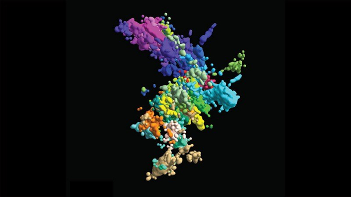 (Xiaowei Zhuang lab)
Above: Multi-coloured image of chromatin, using multiplexed fluorescence in situ hybridisation and super-resolution microscopy.
"It's quite important to determine the 3D organisation," says senior researcher Xiaowei Zhuang, "to understand the molecular mechanisms underlying the organisation and to also understand how this organisation regulates genome function."
Using a new high-resolution 3D imaging system – which involved joining together multiple snapshots of genomic loci along DNA chains – the researchers were able to visualise chromosomes up close in a way that's never been seen before, and even glimpse aspects of transcription activity.
High school and CHEM101 will never be the same. The team is sharing their data online so other researchers can take their analysis further, and so we can explore this (almost) invisible part of ourselves even more in the future.
"We envision broad application of this high-throughput, multi-scale, and multi-modal imaging technology, which provides an integrated view of chromatin organisation in its native structural and functional context," the team explains.
The findings are reported in Cell.