
A new imaging study has found evidence to suggest that malaria parasites gather and accumulate in the spleen, in the first week or so that the infection takes hold.
It's another small win in a string of recent discoveries that are 'redefining' our understanding of malaria – a life-threatening, mosquito-borne disease that affects millions of people worldwide each year.
Australian scientists only recently discovered malaria parasites, especially of the Plasmodium vivax variety, lurking in the spleens of people with chronic malaria infections in "surprisingly large" amounts.
That discovery helps to explain why chronic cases of malaria can fly under the radar on blood tests but suddenly relapse. This new study shows just how fast the parasites find their way to the spleen.
Put together, this research throws weight behind the emerging evidence that the spleen may harbor a reservoir of replicating Plasmodium parasites in malaria. Also, by elucidating another step in the malaria parasite life cycle, it could lead to a rethink of our current disease eradication strategies.
"By performing this novel imaging study in participants undergoing experimental malaria infection, we have been able to look at what is happening inside specific organs during the earliest stages of blood-stage infection," says John Woodford, an infectious disease researcher at the QIMR-Berghofer Medical Research Institute in Australia.
Scientists begun to suspect that P. vivax parasites might fancy the spleen, more so than floating around the bloodstream, because the spleen contains lots of young blood cells, called reticulocytes, that P. vivax can infect.
What they didn't have, until now, was evidence of just how soon Plasmodium parasites might appear in the spleens of people infected with malaria – and if any changes could be detected by imaging, for that matter.
Woodford and his colleagues found evidence of malaria parasites accumulating in the spleen soon after a malaria infection they induced in their volunteers under controlled experimental conditions, and more so with P. vivax than P. falciparum, the deadliest form of Plasmodium parasite that causes malaria in humans.
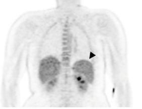 (Woodford et al., PLOS Medicine, 2021)
Above: PET imaging shows increased glucose uptake in the spleen after P. vivax infection.
It's important to note that the study was only small: just seven healthy volunteers, who never had malaria before, were infected with a dose of either P. vivax (for three volunteers) or P. falciparum (the other four) under close observation.
While P. falciparum has been the main focus to date of malaria treatments and eradication efforts, it's P. vivax that is causing a rising number of malaria infections, many chronic and some fatal.
Two types of imaging, MRI and PET, were used to measure the uptake of fluorescently tagged sugar substance in three organs – the spleen, liver, and bone marrow – and the volume of each, roughly a week before and after infection.
Glucose metabolism, a sign of active cells consuming energy, increased within 11 days of infection and this was more pronounced in the P. vivax group, than in those infected with P. falciparum.
Likewise, the volume of these people's spleens showed a similar trend, larger with P. vivax than P. falciparum, but no such changes were seen in the liver or bone marrow.
"This represents further evidence that parasites of the important species P. vivax have a particular predilection for the spleen," Woodford says.
Woodford and his colleagues suggest that the changes in spleen volume and sugar metabolism, could be due to the sheer number of parasites accumulating in the spleen or the activity of the organ itself, in response to a mounting infection – or it could be a combination of both.
"The presence of a hidden compartment [in the spleen] affects our understanding of the basic biology and pathology of this common parasite and may have implications when developing antimalarial treatments," the research team writes in their paper.
Aside from the extra evidence this study provides of Plasmodium parasites in the spleen, it also suggests that whole-body imaging could be a useful tool for studying malaria infections.
"We have demonstrated that the medical imaging techniques MRI and PET may be used to study human P. vivax and P. falciparum infection in life and observe early infection in a way not previously possible," the researchers write.
Before this, scientists have only had blood samples and tissue removed during surgery or after death to rely on. The hope is that perhaps imaging could detect early malaria infections headed for the spleen that blood tests might otherwise miss.
But medical imaging is far more expensive than routine blood tests and might not be readily available in parts of the world where malaria is endemic.
The study was published in PLOS Medicine.