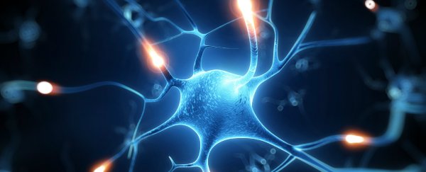
It sounds like something taken straight from a science-fiction movie, but this is science fact: a group of international researchers led by Professor Charles Lieber of Harvard University have developed a method for injecting nano-scale electronic scaffolds into animal bodies. Once connected to electronic devices, these meshes can be used to monitor neural activity and even stimulate tissue and neurons.
Ultimately the methods pioneered by Lieber and his colleagues could lead to new ways to treat neurodegenerative diseases and paralysis, as well as mapping out the brain in greater detail than ever before. Parkinson's is just one of the conditions that could be treated in this way, if it's proved to be safe for humans - so far it has only been tested on mice.
The team brought together by Lieber is made up of internationally renowned physicists, neuroscientists and chemists, and he thinks the technique could make a huge difference in the future. "I do feel that this has the potential to be revolutionary," Lieber said, as Phys.org reports. "This opens up a completely new frontier where we can explore the interface between electronic structures and biology."
"For the past thirty years, people have made incremental improvements in micro-fabrication techniques that have allowed us to make rigid probes smaller and smaller, but no one has addressed this issue - the electronics/cellular interface - at the level at which biology works," adds Lieber. His work has just been published in Nature Nanotechnology.
Once injected, the miniature scaffolding is able to unfurl itself and melds with the existing brain tissue - the neurons apparently look at the new mesh as a friendly support rather than something alien to the body. From there, individual neurons can be both monitored and stimulated through a small connection to the brain. The team says the next step in the research is to try the same technique with larger meshes and more sensors.
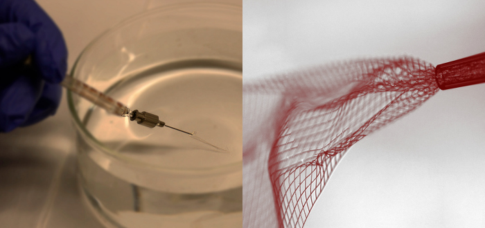 The electronic mesh being injected into a solution unmagnified (left) and through a bright-field microscope (right). Credit: Lieber Research Group, Harvard University
The group of scientists Lieber has brought together are trying to solve a long-standing neuroscience mystery: exactly how the activity of individual brain cells lead to larger cognitive powers (like emotion or perception). Because the new mesh is 95 percent free space, and made of very soft and flexible silk, the brain tissue is able to comfortably rearrange itself around it.
"I think it's great, a very creative new approach to the problem of recording from large number of neurons in the brain," Rafael Yuste, director of the Neurotechnology Centre at Columbia University in New York, told Nature.com.
At this stage not everyone is confident the new procedure can be applied safely to human beings, however. Jens Schouenborg, who is head of the Neuronano Research Centre at Lund University in Sweden, has said he wants to see more evidence of long-term compatibility with the body. Schouenborg is also working on his own gelatin-based 'needle' for delivering electrodes into the brain.