
Just last week, scientists gifted us exquisite images of three of our body's major organs, detailed like never before. From neighborhoods of gut cells to the architects of new life, we saw how cells are arranged and interact.
Now a group of scientists has produced what they claim is the most detailed cell catalogue of the human heart, including the specialized tissue where the heartbeat originates.
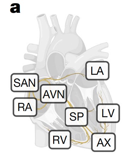
Part of the Human Cell Atlas consortium, which is aiming to map every cell type in the human body, researchers from several UK and German institutes charted eight regions of the human heart, and profiled 75 different cell states that keep the heart moving and help defend it from infections.
The map isn't something most of us can visually appreciate. It's more like a molecular catalogue of cell types and their active genes, and it could help understand diseases such as those that affect heart rhythm.
As we can feel inside our pounding chests, the heart is muscle in motion and electrical impulses in action. Heart contractions result from the collective motion of heart muscle cells, sparked by electrical impulses in so-called pacemaker cells.
These pacemaker cells are mostly found in the sinoatrial node of the heart, one part of the cardiac conduction system that includes a few other interconnected nodes and cell bundles, which scientists don't fully understand.
"The cardiac conduction system is critical for the regular and coordinated beating of our hearts," explains James Cranley, a cardiologist specializing in heart rhythm disorders and joint lead author of the study. "Yet the cells which make it up are poorly understood."
To resolve those cell types in greater detail, Cranley and colleagues used single-cell transcriptomics methods, which decipher how the genetic instructions encoded in DNA are read in individual cells.
They applied these methods to tissue samples from 25 donor hearts that were not quite suitable for organ transplantation, but invaluable for this study, which analyzed more than 700,000 individual cells and nuclei.
By mapping out distinct clusters of heart cells in multiple donors who were otherwise healthy, the team discovered pacemaker cells in close connection to glial cells.
Glia usually support neurons in the brain and wider nervous system. But in the sinoatrial and atrioventricular nodes, and atrioventricular bundle of the heart, the researchers found glial cells supporting signaling processes in pacemaker cells.
The pacemaker cells were 'enveloped' in the spindly extensions of glial cells, their connections resembling how nerve cells come together at synapses.
The researchers also surveyed the outer layer of the donor hearts. There they found immune cells called plasma cells and confirmed that they produce antibodies to shield the heart from infections in the nearby lungs.
With the myocardium, the muscular tissue of the heart, Cranley and colleagues identified a population of cells that appear particularly sensitive to stress and inflammation.
The cells had lots of genes encoding receptors for inflammatory signaling molecules, and expressed high levels of a peptide that has been linked to heart failure.
"By understanding these cells at an individual genetic level, we can potentially develop new ways to improve heart treatments," says joint lead author Kazumasa Kanemaru, a cardiac genomics researcher at the Wellcome Sanger Institute in the UK.
Moreover, the researchers categorized pacemaker cells based on the types of ion channels they express, hoping their findings can further research into what happens when the heart's wiring system misfires, and why some heart therapies fail to work as designed.
Ion channels are cellular gatekeepers that let charged molecules flood into and out of cells. This brief polarization triggers all-important electrical signals in pacemaker cells that jump-start the heart.
"Altogether, these data provide a highly specific map of genes and cells of the [cardiac] conduction system," the researchers conclude.
The study has been published in Nature.