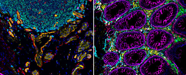
A consortium of scientists has just published an atlas of remarkable images of three human organs, each vital in their own way, showing how cell types are arranged and interact.
The result: Glittering, kaleidoscopic blueprints lit up by fluorescent dyes that reveal new intimacies about our bodies and reshape our understanding of human biology and disease like never before.
As you can see in the diagram below, researchers generated the cell atlases in three ways.
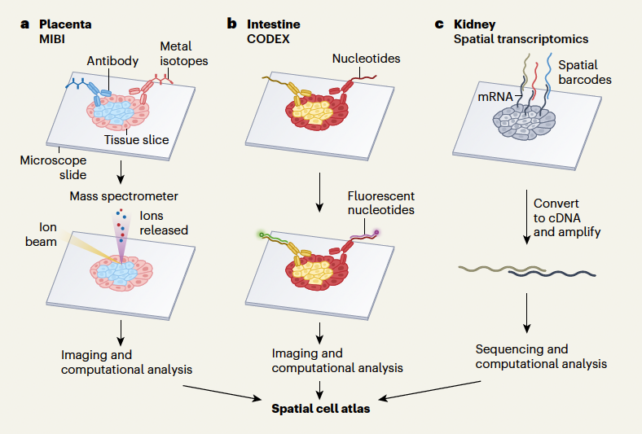
One team, led by Washington University nephrologist Sanjay Jain, used single-cell transcriptomics methods, which reveal how the genetic instructions encoded in DNA are read in individual cells, to map the kidney.
Another group headed up by genomicist Michael Snyder at Stanford School of Medicine mapped the intestine with fluorescent antibodies bound to tissue sections, imaged under the microscope.
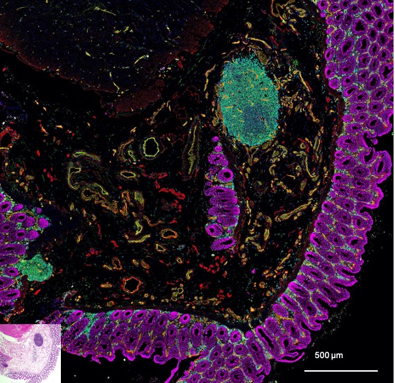
And a third team peered into what scientists have described as "arguably the most important organ of the body, but paradoxically the most poorly understood" – the placenta.
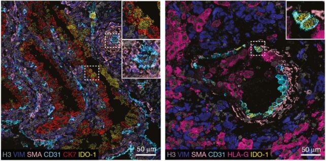
Stanford University pathologist Michael Angelo and colleagues imaged slices of placental tissue treated with metal ions chemically linked to antibodies capable of latching onto signature compounds on the surfaces of cells, focusing on samples where placental cells had attached to the wall of the uterus.
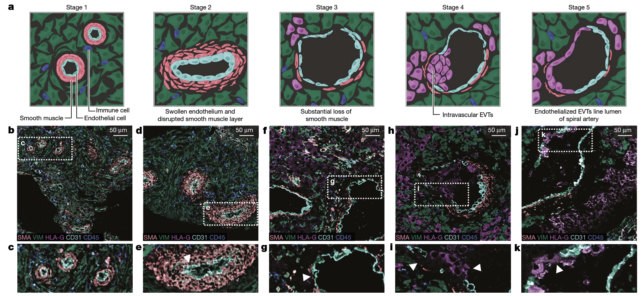
By imaging multiple samples at different stages of this process, from 6 to 20 weeks' gestation, researchers plotted interactions between placental cells and the mother's immune cells and arteries – both of which adjust to accommodate the placenta.
We can see, in exquisite detail, how this remodeling process "allows peaceful coexistence between genetically distinct maternal uterine and fetal placental cells," according to two cell biologists at the Wellcome Sanger Institute, Roser Vento-Tormo and Roser Vilarrasa-Blasi, who penned a commentary about the collection of new papers.
As for the intestine, this meters-long organ is where millions of microbes jostle about, ultra-processed foods trigger inflammation, and cells are plugged into the body's 'second brain'.
Snyder's team discovered drastic shifts in how cells were arranged along the length of the intestine. They sketched out distinct neighborhoods stacked with immune cells ready to launch into action and stumbled upon new subtypes of epithelial cells that line the gut.
Further imaging of the ruffled surfaced of the intestine and its layers could reveal new insights about how inflammatory bowel diseases, mood disorders, or even neurodegenerative diseases develop.
Kidneys, too, do so much for the body. They pump blood to clean it of toxins and waste products, but often fail or become diseased and need replacing.
Sampling more than 90 kidneys, Jain and colleagues outlined communication channels between cells and located cells in which repair pathways become defective during acute kidney injury or chronic kidney disease.
"We looked at how kidney cells are organized, their molecular identities, and how they shift from healthy to diseased states," explains Jain, who led the kidney imaging study.
"With this knowledge, we can start thinking about the drugs or small molecule targets that can prevent progression to disease or promote recovery from injury."
Bear in mind these images were taken using a small number of precious samples generously donated by people undergoing surgery and volunteering to participate in research.
A much harder task will be appreciating the structural differences in organs that exist between different groups and populations that can have real health consequences, even when we're all made to roughly the same body plan.
The papers and the accompanying commentary have been published together in Nature.