For the first time ever, scientists have managed to detect a single atom using super high-resolution magnetic resonance imaging.
Researchers from the Laboratory for Solid State Physics at ETH Zurich in Switzerland have built a tiny magnetic resonance imaging (MRI) machine that's powerful enough to detect a single hydrogen atom - a huge breakthrough in the quest to make MRI more powerful.
Currently, MRI technology used in hospitals works by creating a powerful magnetic field with electromagnetic coils, and then measuring how the nuclei of the hydrogen atom in our tissues respond. This gives doctors an image of the composition of different types of tissues and allows them to spot objects as small as one-tenth of a millimetre, such as tumours.
But while this is great to help diagnose disease, an important goal for scientists is to be able to image single molecules - something that would require more than one million times finer resolution than is currently available.
By detecting the signal from a single hydrogen atom, scientists have now taken an important step towards that goal. This new nano-MRI technology could be used to help scientists image individual proteins and find out more about their structures, something that could be incredibly useful for drug development.
"MRI is nowadays a mature technology and its spatial resolution has remained largely the same over the last ten years. Physical constraints preclude increasing the resolution much further," said Christian Degen, the research leader, in a press release.
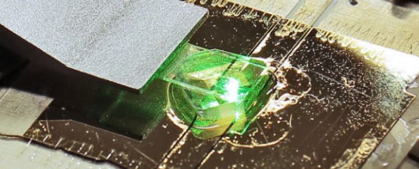
To create such high-resolution MRI technology, the scientists moved away from relying on the electromagnetic coil that's used in hospitals. Instead, the team used a diamond sensor chip to measure the magnetisation of atoms. The chip was embedded into a fluorescence microscope, which then provided an optical readout of the results.
This sensor chip that they created contains a diamond that has an impurity in it - this means there are two carbon atoms missing from the regular diamond lattice, and one of these has been replaced with a nitrogen atom.
This creates what is called a nitrogen-vacancy centre in the diamond, an impurity that is both fluorescent and magnetic, which means it's able to measure very small changes in magnetic fields.
For this experiment, the scientists used a piece of diamond that had the nitrogen-vacancy centre just a few nanometres below the surface, and covered it in a thin layer of inorganic salt. The diamond was able to confirm the presence of other magnetic atomic nuclei in the immediate vicinity, and using quantum mechanics, the team were able to work out whether they'd detected an individual nucleus, or a cluster of several hydrogen atoms.
Not only that, but using the data collected from the diamond, the scientists were able to work out where the hydrogen nuclei was with respect to the nitrogen-vacancy centre with an accuracy of one-ten-millionth of a millimetre. Their results are published in Science.
"This is an important intermediate step toward the mapping of entire molecules," said Degen in the release.
The next step is to image a small molecule with the nano-MRI device.
Right now scientists study the structure of molecules using X-ray crystallography, but this requires growing crystals consisting of billions of identical molecules, something that's difficult and often impossible with proteins.
If this nano-MRI device is successful at measuring molecules, not only will it help scientists understand the structure of proteins, it will also allow them to label proteins with isotopes and monitor their function throughout the body.
Sources: Futurity, ETH Zurich