For the first time, scientists have been able to accurately measure the mass of the human chromosome.
Using a powerful X-ray source at the UK's national synchrotron science facility, the Diamond Light Source, physicists were able to determine the individual masses of all 46 chromosomes in human cells.
The masses were found to be significantly higher than expected – around 20 times higher than the DNA contained therein – likely reflecting the additional mass of other unknown elements inside chromosomes that we are yet to discover, the researchers suggest.
"The mass of DNA we know from the Human Genome Project, but this is the first time we have been able to precisely measure the masses of chromosomes that include this DNA," said biophysicist Ian Robinson of University College London.
"Our measurement suggests the 46 chromosomes in each of our cells weigh 242 picograms (trillionths of a gram). This is heavier than we would expect, and, if replicated, points to unexplained excess mass in chromosomes."
Chromosomes are little thread-like packages of DNA that can be found in the cell nuclei of living organisms. Each chromosome contains one molecule of DNA, which in turn contains the genetic instructions for the development and life of that organism.
Humans have 23 pairs of chromosomes, representing 22 pairs of numbered chromosomes (autosomes), and one pair of sex chromosomes.
Chromosomes keep the DNA inside from unravelling, helping to maintain its structure during the cell replication process.
Chromosomes were first discovered in the 19th century, and since that time scientists have learnt a lot about their role in keeping living organisms functional. There is, however, a lot we still don't understand. In this case, scientists used a technique called hard X-ray ptychography to probe inside them.
This involves using a type of particle accelerator called a synchrotron to produce a powerful beam of X-rays. When these X-rays pass through the chromosomes, their diffraction creates an interference pattern that scientists can use to create a high-resolution 3D reconstruction of that chromosome.
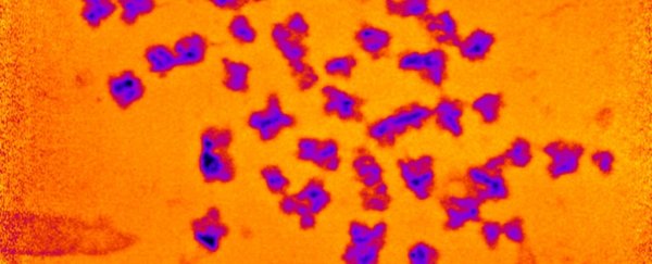
The researchers imaged human white blood cells at metaphase (a phase in the cell cycle in which chromosomes are condensing), and just prior to cell division, when the 46 chromosomes inside each cell have tightly packaged up the DNA inside.
Using this technique, the researchers were able to determine the number of electrons, or electron density, in the chromosome. The mass of electrons is well known; in fact, the rest mass of electrons is one of the fundamental physical constants. So, the research team was able to use this to calculate the mass of the chromosome.
It's not entirely clear what might be accounting for the unexpected mass the researchers found, but the insights here could have untold benefits for science, helping us towards a better understanding of how our bodies work, and why things sometimes go wrong.
"A better understanding of chromosomes may have important implications for human health," said bioscientist Archana Bhartiya of University College London.
"A vast amount of study of chromosomes is undertaken in medical labs to diagnose cancer from patient samples. Any improvements in our abilities to image chromosomes would therefore be highly valuable."
The research has been published in Chromosome Research.