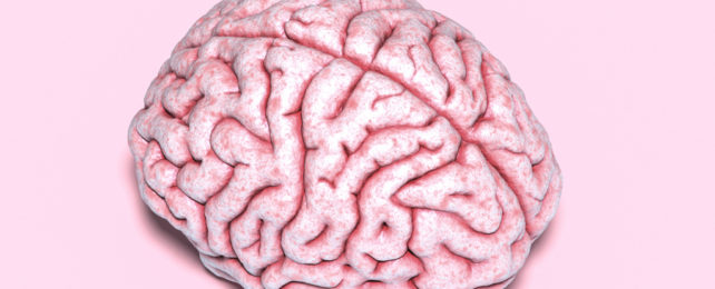
The folds of the human brain are instantly recognizable. Snaking ridges and deep furrows give the squishy tissue inside our heads structure and the appearance of a wrinkly walnut.
Into peaks called gyri and fissures called sulci, the outermost layer of brain tissue is folded so that reams of it can be squeezed into the skull, and it's here, on the brain's wrinkly surface, that memory, thinking, learning, and reasoning all happens.
This folding, or gyrification, is crucial for proper brain function and circuitry – and is said to be why humans have greater cognitive abilities than apes and elephants, whose brains have some folds, and rats and mice, whose smoothed-surfaced brains have none.
Now, a team of scientists has discovered why some people have more brain folds than others, in a condition that affects normal brain development called polymicrogyria (PMG).
In polymicrogyria, too many gyri are piled on top of each other, resulting in an abnormally thick cortex and leading to a broad spectrum of problems such as neurodevelopmental delay, intellectual disability, speech difficulties, and epileptic seizures.
"Until recently, most hospitals treating patients with this condition did not test for genetic causes," explains University of California San Diego (UCSD) neuroscientist Joseph Gleeson, one of the researchers behind the new study.
Polymicrogyria comes in many forms, with localized or widespread cortical thickening detectable on brain scans.
Mutations in 30 genes and counting have been associated with the condition. But how any of those genetic errors, alone or in tandem, result in the overfolded brain tissue remains unclear. Many cases of PMG also lack an identifiable genetic cause.
It's thought to have something to do with the tardy migration of cortical brain cells in early development that leads to a disordered cortex. The cortex is the outermost layer of the brain's two-lobed cerebrum, a thin sheet of gray matter composed of billions of cells.
To investigate further, Gleeson collaborated with researchers at the Human Genetics and Genome Research Institute in Cairo to tap into a database of nearly 10,000 families from the Middle East affected by some form of pediatric brain disease.
They found four families with a nearly identical form of PMG, all harboring mutations in one gene. That gene encodes a protein that clings to the surface of cells, with the imaginative name of transmembrane protein 161B (TMEM161B). But no one knew what it did.
Gleeson and colleagues showed in subsequent experiments that TMEM161B is found in most fetal brain cell types: in progenitor cells that grow into specialized neurons, in mature neurons that excite or inhibit their neighbors, and in glial cells that support and protect neurons in various ways.
However, TMEM161B is from a family of proteins that first appeared, evolutionarily speaking, in sponges – which have no brain.
This puzzled Gleeson and fellow UCSD neuroscientist Lu Wang who wondered if the protein might indirectly affect cortical folding by meddling with some basic cellular properties that give shape to complex tissues.
"Once we identified TMEM161B as the cause, we set out to understand how excessive folding occurs," says Wang, the study's lead author.
Using stem cells derived from patient skin samples, the researchers generated organoids, tiny tissue replicas that self-organize in plastic dishes the way bodily tissues and organs do. But the organoids made from patient cells were highly disorganized and showed disrupted radial glial fibers.
In the developing brain, these progenitor cells – which give rise to neurons and glia – usually position themselves at the apex of the cortex and extend radially downwards towards the bottom layer of cortical tissue. This creates a scaffolding system that supports the migration of other newly formed cells as the cortex expands.
But without TMEM161B, radial glial fibers in the organoids had lost sense of which way to orient themselves. Further experiments also showed that the cells' internal cytoskeleton was a mess.
So it seems that without their own internal scaffold, radial glial fibers cannot be the scaffold other cells need to find their way into position in the developing brain.
While this discovery is a promising step forward, giving us clues to how the condition unfolds, it may only be relevant to a small or as yet unknown fraction of PMG cases.
Much more research is needed to flesh out our understanding of how many people with PMG are affected by mutations in TMEM161B – but now researchers know what to look for, they can trawl other datasets looking for more cases.
"We hope that physicians and scientists can expand upon our results to improve diagnosis and care of patients with brain disease," says Gleeson. That's a long road but a hopeful one.
The study has been published in PNAS.