You're piloted through the world by your subtalar joint, sometimes thought of as the 'steering wheel' of the body. Now, for the first time scientists have taken a detailed look at the way this crucial piece of our anatomy works - which could help us to better treat ankle injuries one day.
Before now, the joint had never been imaged while rotating in a standing position (bearing the whole weight of the body), which makes it difficult to diagnose and treat ankle problems, including the classic sprained ankle. If doctors don't know what's happening, they're less able to help.
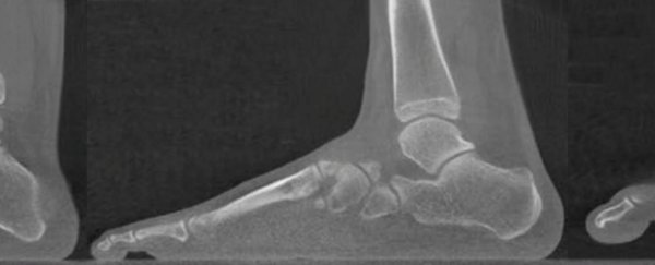
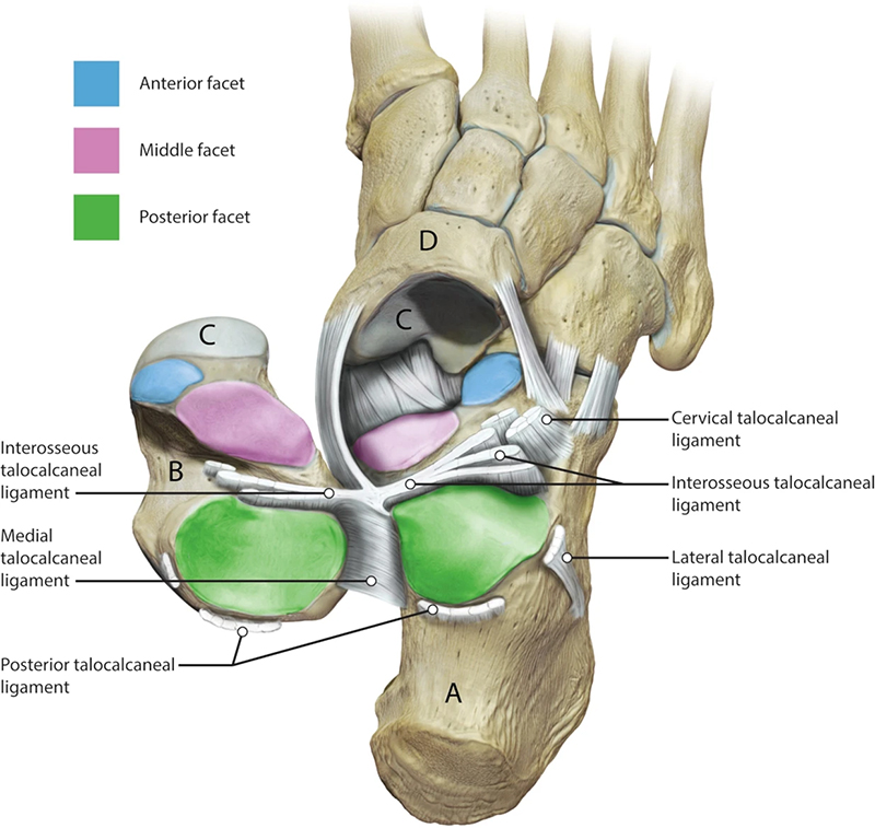
The subtalar joint sits below the ankle, and is made up of two bones: the calcaneus (heel bone, labelled A in the diagram below) and the talus (labelled B).
The joint is key to some pretty complex mechanisms – keeping the ankle flexible for twisting and bending, but rigid for propelling the body forward. The whole system is held in place by a bunch of ligaments.
 (Fernández et al, Scientific Reports)
The new imaging was done through a sophisticated 3D technique using computed tomography (CT scans) and something called digital volume correlation, where several images are combined to produce an accurate model of movement.
"This is the first time this technique has been used in humans," says bioengineer Gianluca Tozzi, from the University of Portsmouth in the UK.
"It is non-invasive and gives clinicians a perfect view of a patient's subtalar joint motion under full weight-bearing, making it possible for the first time to determine the joint's centre of rotation."
Getting a close-up view of how this works can be very useful for all kinds of reasons, from improving the diagnosis of injuries to the ankle, to developing replacement prosthetic parts when wear and tear takes its toll.
Doctors can't currently replace your subtalar joint in the same way they can your hip, for example – and that might change now we can actually see how the subtalar joint works inside the body.
It's also hoped that it might lead to more tailored treatments for ankle problems, like the personalised ones currently prescribed for issues with hips and knees.
Problems with the subtalar joint can have a significant negative impact on day-to-day activities as well as anything (like sports) that requires a lot of movement. In the elderly, osteoarthritis in this region can be as bad as end-stage heart disease in terms of how much it affects quality of life.
Looking further ahead, the technique that the scientists have developed in the lab – and tested on eight volunteers – could be expanded to look at other joints and other types of strain.
"I've always hoped for this," says Tozzi. "Everyone working in healthcare research hopes their work will be transferable from the lab to real life, making a difference to patients."
"This new research helps us to better understand the complex biomechanics of the foot and could pave the way for new treatments that just aren't currently available."
The research has been published in Scientific Reports.