Anyone familiar with the appearance of the human brain would recognise the distinctive, wrinkly folds that cover its surface. Scientists believe that the brain's folding enables a large cortex to fit into a smaller volume – reducing wiring length and improving cognitive function – but how does the brain physically end up in this convoluted shape?
Researchers at Harvard University have gotten closer to understanding how this process works, building a 3D-printed layered brain model that replicates the manner in which real brain cortices fold in upon themselves.
Brain folding doesn't actually appear in all animals, and is limited to a number of species including some primates, dolphins, elephants, and pigs. While the link between these animals' brain folding and their comparatively high cognitive functions has been noted before, it is not yet fully understood by scientists.
In humans, the folding starts at about the 20th week of gestation of a foetal brain, with the process continuing until a child is approximately 18 months old.
The folding happens as the brain grows, with the number, size, shape, and position of neuronal cells all contributing to the expansion of the cortex – also called grey matter – relative to the white matter that lies underneath.
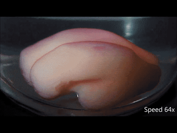
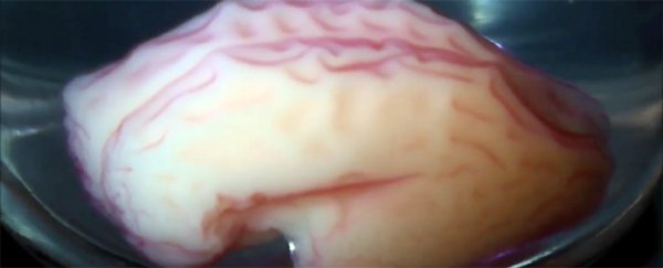
To mimic this folding motion with an artificial brain structure, the researchers sourced MRI images of human foetuses. With the data in hand, they made a 3D gel model of a smooth, unwrinkled foetal brain as it would look before any of the folding takes shape.
The model's surface was then coated with a thin layer of elastomer gel, effectively representing an artificial cortex. To replicate the natural process of cortical expansion, the gel brain was immersed in a solvent, causing the outer layer to swell and expand. Within minutes – sped up considerably in the GIF seen here – the artificial brain's outer layer resembles the formation of folds in real brains.
"We found that we could mimic cortical folding using a very simple physical principle and get results qualitatively similar to what we see in real foetal brains," said one of the researchers, L. Mahadevan. "This simple evolutionary innovation, with iterations and variations, allows for the thin but expansive cortex to be packed into a small volume, and is the dominant cause behind brain folding, known as gyrification."
According to the researchers, the shape and position of the folds that result from the cortex expansion are critical to health, with the form of the brain related to its function.
"The geometry of the brain is really important because it serves to orient the folds in certain directions," said one of the researchers, Jun Young Chung. "Brains are not exactly the same from one human to another, but we should all have the same major folds in order to be healthy."
The findings, which are reported in Nature Physics, could help scientists to better understand how the outer shape of the brain is related to its inner workings.
"Our research shows that if a part of the brain does not grow properly, or if the global geometry is disrupted, we may not have the major folds in the right place, which may cause potential dysfunction," said Chung.
