Thousands of years ago, near what is today the British village of Heslington, a man's body started to decompose. Flesh and organs became mud. Hair turned to dust. In the end, bones remained, and, mysteriously, a small piece of his brain.
After months of patiently investigating the tissue's proteins, an international team of researchers finally has clues explaining this remarkable instance of preservation, and it could help us better understand how healthy (and unhealthy) brains actually work.
The 2008 discovery of the Heslington brain – one of the oldest specimens of human neural tissue ever to be uncovered in the UK – left researchers with a challenging puzzle to solve.
Within moments of a typical death, brain tissue starts to decompose. Compared with other body parts, this decay is especially rapid, with various proteins going to work demolishing cellular infrastructure.
So when archaeologists looked inside a mud-caked skull pulled from an Iron Age dig site, they were understandably shocked to see the withered remains of what looked like a chunk of recognisable human brain.
According to carbon dating, the middle-aged man breathed his last breath somewhere between 673 and 482 BCE, most likely as the result of a fractured spine – the kind you get after a hanging.
Exactly who he was, or why he died, probably won't ever be known. Sometime after his speculated execution, though, the victim's severed head was thrown into a pit, where it was encased in a fine grain sediment.
Soft tissues can often be preserved if they're desiccated, frozen, or kept in an anaerobic, acidic environment.
What's especially strange in the case of the Heslington skull is the lack of preservation of any other part of the body, including hair.
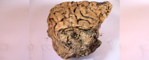
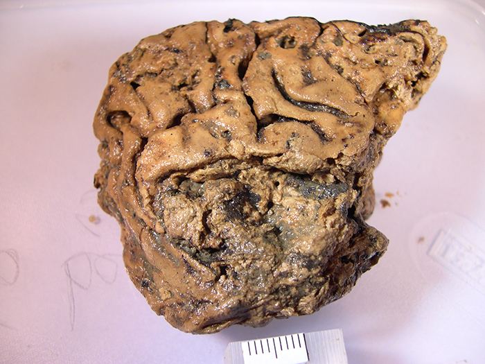
For all appearances, the firm, tofu-like material looks like a caramelised chunk of human cerebral cortex, only it's 80 percent smaller than an adult human brain.
 The Heslington brain. (Dr Axel Petzold)
To work out what made the remaining organic material so special, researchers took a closer look at the nature of its proteins.
Unlike most organs, the brain needs to be well supported on a cellular level to operate, maintaining connections within the complex weave of neurons and their long bodies.
A matrix of intermediate filaments (IFs) performs this task in living brains, and it seems under the right circumstances, they can retain some kind of integrity long after the cells have been reduced to molecular ashes.
We already know a fair bit about these IFs based on various pathological studies. Different cell types have their own types of filament, and this specificity has attracted research for uncovering biomarkers for neurological diseases.
In the case of the Heslington brain, microscopy revealed weaves of IFs that resembled the long threads of axons making up a living brain, only shorter and narrower, while antibody markers matching axon proteins confirmed they once housed the long neuron tails.
Further analysis with specific antibody markers revealed a disproportionate amount of neural structures belonging to 'helper' cells such as astrocytes, with fewer proteins marking out thinking grey matter tissue.
Determining why these particular astrocyte IFs in particular didn't follow the usual path of decay was never going to be simple.
There were no signs of the preserving tannins often seen in British bog bodies, and while the specimen's pH was towards the lower end, the researchers weren't confident they could use it to estimate the acidity of the body's grave.
What's more, proteins that stick around at relatively warm temperatures tend to form stable structures, and stable proteins don't unfold as easily as unstable ones.
So over the course of a year, the researchers patiently measured the slow unwinding and breakdown of proteins in a modern specimen of neural tissue and compared it with the decay within the Heslington brain.
The results invited speculation over a chemical that blocks destructive enzymes called proteases in the months following death, allowing the proteins to coalesce into stable aggregates that could persist at warmer temperatures.
"Combined, the data suggest that the proteases of the ancient brain might have been inhibited by an unknown compound which had diffused from the outside of the brain to the deeper structures," they write in their report.
What seems clear is that there was nothing particularly special about this poor Iron Age fellow's brain. Rather, something in the environment could have inhibited the chemical processes that would ordinarily break down the protein filaments responsible for supporting the brain's 'white matter' astrocytes, at least long enough for it to clump into a more robust form.
Of course, with only this incredibly unique sample to study, it's hard to draw firm conclusions.
But even if the proposed 'unknown blocker' turns out to be a red herring, research on the way that IFs form stable aggregates could inform models explaining how destructive plaques form in our brain.
And with possible scraps of protein being found in fossils from time to time, it would be good to have a sound understanding of how they might 'unfold' to deduce their original structures.
The strange brain from Heslington still has a few things to teach us yet.
This research was published in Interface.
A version of this article was first published in January 2020.