'Painting' with fluorescent markers has long been a handy way to detect unique double-stranded structures in DNA. Once limited to a palette of just 256 colors, scientists can now create stunning works of laboratory art with an incredible 16 million shades and hues.
The new technique accurately recreates digital images with 24-bit color depth, and the results are breathtaking.
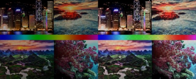
Here's an original digital image:
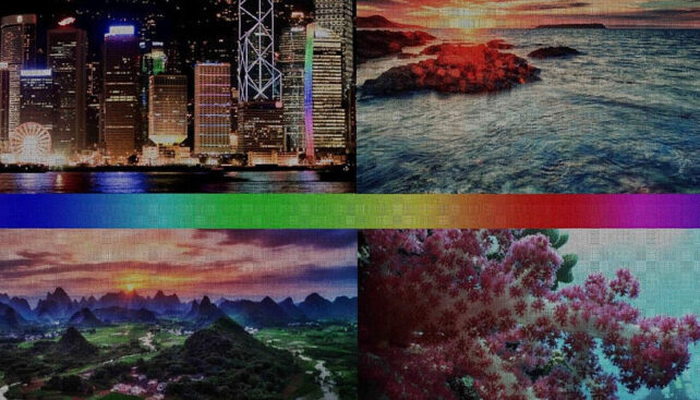
And the DNA 'painted' version:

It's not just a new artistic format. The miniaturized DNA-based painting approach is a scale-up on microarray technology for studying gene expression, and it has other potential too.
Researchers often use works of art to test or showcase techniques that may later prove useful in real-world applications.
"Beyond imaging, a DNA color code could have very useful applications in data storage on DNA," says chemist Tadija Kekić from the University of Vienna.
Converting data into DNA sequences stored on a chip is similar to storing information in a barcode. This new method by Kekić and another University of Vienna chemist, Jory Lietard, could allow a greater amount of storage on a smaller surface.
Their technique is so precise it could be used to paint micrometer-scale features onto biopolymers. Possible uses include biosensors and diagnostics, where fine-tuned control of DNA self-assembly is crucial.
A huge amount of information can be stored in DNA as a code consisting of sequences made up of four chemical bases – adenine, guanine, cytosine, and thymine. Each base also corresponds to a partner, seeing to the formation of complementary sequences in the form of a double strand.
Having one sequence in hand gives scientists a tool for finding its matching partner in a jumbled mess. Analytical techniques based on DNA sequences stuck to a solid surface in a grid pattern, called DNA microarrays, rely on colored fluorescent markers to show when complementary DNA strands bind together.
This process where complementary DNA strands recognize each other and bind together as double strands is known as hybridization. Living organisms use the simple, stable rules of hybridization to read and copy their genetic information.
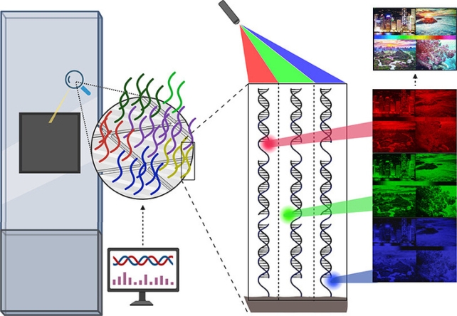
To visualize how we can turn fluorescent DNA sequences into vibrant works of art, it might help to picture the way modern color displays, like phones and laptop monitors, create a wide range of colors.
The color of each pixel in the grid of your screen is made up of red, green and blue primary channels, and the intensity of each channel can be dialed up or down to produce the color you see.
Having mismatches between bases on a double strand essentially adds a small amount of instability to its hybridization, changing the molecular structure of double-stranded DNA.
The researchers behind this new fluorescent hybridization technique programmed instability into the strands to change the brightness of their fluorescent markers. By using different dyes for different colors and removing bases, hybridization on patterned DNA surfaces can make patterns that are really easy to see.
Specific dyes (Cy3, Cy5, and fluorescein) on fragments of DNA, called probes, create 256 shades of light in each of the red, green, and blue channels. This creates a slider-like fluorescence signal between none of the color and full color.
"Essentially, our synthesis surface becomes a canvas for painting with DNA molecules at the micrometer scale," says Lietard.
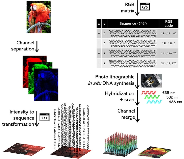
To demonstrate the color range, the team separated digital images into three 8-bit RGB layers and assigned DNA sequences for each pixel's value. On a fingernail-sized microarray, the technique – maskless array synthesis combined with photolithography – can synthesize hundreds of thousands of unique DNA sequences at once.
With the help of a device with hundreds of thousands of tiny mirrors corresponding to the pixels in the image, and computer scripts, more than 786,000 DNA sequences can fit on the microarray surface in a grid formation. That's one for each pixel unit of 14 x 14 micrometers in the RGB layer, with information on the intensity value encoded in the DNA segment.
Scanning the three tiny microarrays, or precisely arranged 'DNA canvases', and then merging the layers back together digitally, allows reproduction of the original image in more than 16 million colors with a resolution of 1024 x 768 pixels. The team thinks the process could be expanded to work at full HD and even 4K.
"Digital images can be reproduced with high fidelity at the micrometer scale by using a simple process," the authors write.
Among many uses, higher resolution fluorescence signals could lead to more precise measurements of processes occurring inside our bodies, allowing better understanding of cell biology, and earlier detection of diseases like cancer.
The study has been published in the Journal of the American Chemical Society.