Devoid of a head, many insects will continue to kick and twitch until at last, drained of all life, their movements grind to a complete stop.
Scientists have known for some time that the spinal cord is capable of executing limb movements beyond reflex jerking motions, even to the point of adapting to avoid unpleasant stimulations.
Just how its neurons 'learn' new responses without the brain's say-so has never been clear.
A study on transgenic mice conducted by researchers from the VIB-Neuro-Electronics Research Flanders in Belgium has discovered the role of a specific gene expressed in spinal nerves in memorizing responses to potential threats.
"Not only do these results challenge the prevailing notion that motor learning and memory are solely confined to brain circuits, but we showed that we could manipulate spinal cord motor recall, which has implications for therapies designed to improve recovery after spinal cord damage," says neurologist and senior researcher Aya Takeoka.
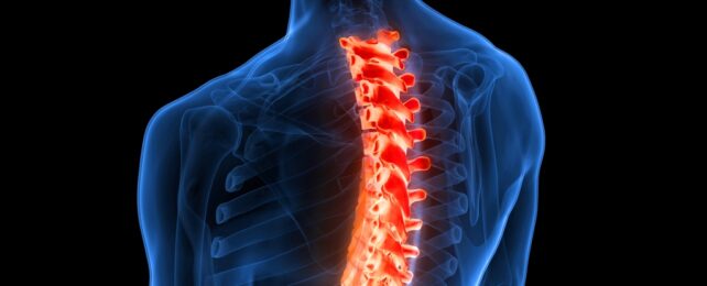
While the brain has the ultimate call over most forms of movement, the spinal cord is more than just a simple highway for nervous signals. It contains genetically diverse populations of neurons capable of molding to suit individual needs in locomotion or withdrawal from pain as the individual develops.
As complex as the organ is, the nerves in the spinal cord can be divided broadly into two basic categories – those that carry sensory information, called dorsal neurons, and tissues that control motor responses, called ventral neurons.
Within each class, inhibitory neurons act as boom-gates, fine-tuning and coordinating sensations and movements on the brain's behalf.
How these distinct classifications of spinal cord tissue work together to learn novel responses long after the nerves have locked in place has been a pressing question for neurologists seeking ways of helping damaged nerves recover.
Takeoka and her team placed mice with transected spinal cords in harnesses that suspended their hind limbs in the air, allowing them to move freely. Without their brains sending and receiving signals from their back legs, all responses were left to their spinal nerves.
By stimulating the test animals' feet with mild zaps of electricity – one at random times, the other only in response to a set amount of leg drooping – the researchers were able to test whether the spinal cord could learn how to react to a negative stimulus.
With one mouse learning to pull up its hind legs, the team switched the roles for each mouse a day later, demonstrating the learned responses weren't short-term adaptations. The nerves had truly learned a new trick.
The team then used six different kinds of genetically altered mice to single out the likely mechanisms that preserved the memory of the electrical shock in the spinal nerves.
Excluding different types of genetically distinct nerve cells one by one, they found those with impeded nerves at the top of the cord, especially those lacking a functional Ptf1a gene, were incapable of adapting to the shocks. Among those that had adapted, disabling Ptf1a did not reverse what they'd learned.
But switching off a second gene encoding for homeobox protein engrailed-1 (En1 gene) in the ventral nerves towards the lower part of the spinal cord effectively made the adapted mice 'forget' how to respond to shocks in follow-up tests a day later. Exciting those same nerves artificially, on the other hand, returned their ability to recall the reflex.
From a medical point of view, understanding how our spinal cord can remain plastic throughout life and continue to respond to environmental changes could inspire novel research into treatments for nervous system damage in humans.
"Gaining insights into the underlying mechanism is essential if we want to understand the foundations of movement automaticity in healthy people and use this knowledge to improve recovery after spinal cord injury," says Takeoka.
This research was published in Science Advances.