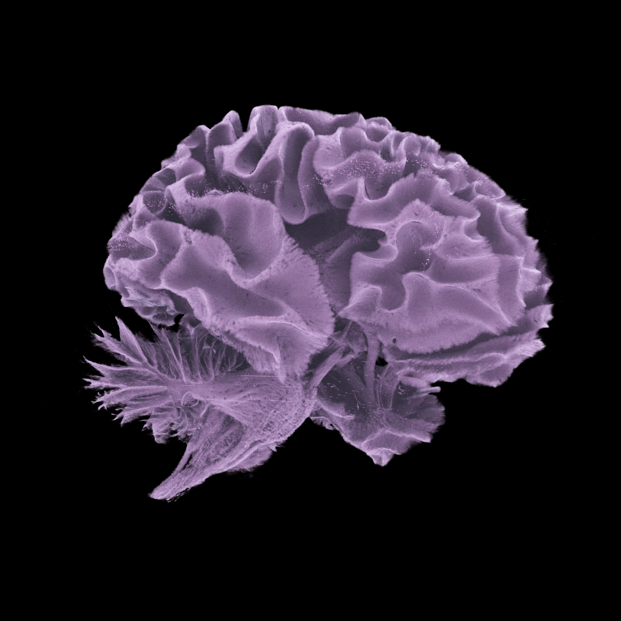
A pioneering project has revealed the human body like never before, from entire organs down to cellular structures, with unprecedented precision on the scale of a single micron – about 50 times thinner than a strand of human hair.
Our bodies are akin to biological nesting dolls, formed from a hierarchical assembly of cells, tissues, and organs whose structure underpins how they function, interact, and respond to disease.
Now, the Human Organ Atlas (HOA) provides a "new gold standard" in medical imaging by showing the framework of the brain, heart, lungs, liver, kidneys, and other bodily systems in stunning detail and three dimensions.
The HOA aims to democratize vast amounts of scientific data, including numerous images exceeding one terabyte (TB). For perspective, one TB is equivalent to more than 250,000 photographs, 17,000 hours of audio files, or 85 million Microsoft Word documents.
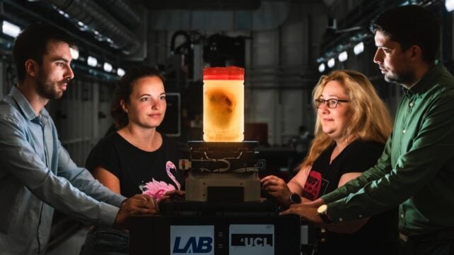
"This is a resource for researchers, doctors, educators – but also for anyone curious about how the human body is built," says Paul Tafforeau, the beamline scientist at the European Synchrotron Radiation Facility (ESRF) in France who pioneered the advanced imaging method used to build the HOA.
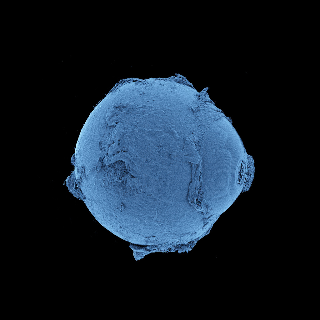
Called hierarchical phase-contrast tomography (HiP-CT), the technique uses X-rays generated by high-energy particles zipping inside a synchrotron, or particle accelerator, called the Extremely Brilliant Source (EBS).
This fourth-generation accelerator provides a medical imaging tool that is up to 100 trillion times brighter than regular hospital X-rays.
HOA researchers have so far used the EBS to non-destructively image intact, ex vivo organs from dozens of donors, achieving a unique level of zoom with cellular resolution.
"To create the Human Organ Atlas, we brought together scientists and medics from nine institutes worldwide," explains materials scientist Peter Lee, of University College London (UCL).
"This grouping is continuing to expand, helping gain new insights into diseases from osteoarthritis to heart disease and changing how we learn about the human body."
HiP-CT imaging has, in earlier studies, revealed previously unknown disease pathways at microscopic scales, including vascular damage in the lungs of individuals who died from COVID-19, as well as vascular features of adenomyosis, a non-cancerous gynecological disorder.
At the time of writing, the continuously updated HOA includes 87 organs and 363 three-dimensional datasets, made possible by 54 donors thus far.
In some cases, the HOA contains images of multiple organs from a single donor, including one individual who had a history of high blood pressure, allowing clinicians to analyze the impact across different organ systems – a primary research goal.
Various other ailments are captured, including cancer, which is among the leading causes of death in the Global North, as well as rare pathologies, such as Dandy-Walker syndrome, a congenital condition affecting fewer than 1 in 30,000 newborns.
Aside from medical training and education, the HOA could also be used to train machine learning models, which are becoming increasingly common in healthcare, for better or for worse.
Using such a comprehensive, high-resolution training set to train AI may lead to improved disease detection and more effective treatment strategies.
"I am personally hugely excited to see how the AI community [uses] the Human Organ Atlas in AI foundation models," says UCL biophysicist Claire Walsh, director of the HOA Hub.
By illuminating the unplumbed depths of human physiology, the researchers hope their work will improve public science engagement, with additional advances on the horizon.
Related: Scientists Beamed Light Right Through a Man's Head For The First Time
"Currently we work on isolated organs, but in the future, we expect to develop the technique to be able to image complete human bodies with a resolution 10 to 20 times higher than what is possible today," Tafforeau says.
"Such data could transform how anatomy is studied and understood."
This research was published in Science Advances.