If you could peer inside a cell's nucleus, you would find DNA tightly packed into a remarkably small space.
But that arrangement presents a puzzle: DNA molecules carry the same negative electrical charge, so they repel one another. How can they get so close?
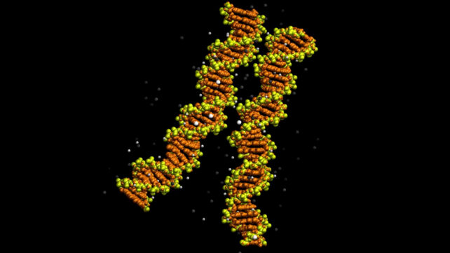
Researchers at the University of Sheffield and the University of York in the UK have captured a striking clue. Two DNA double helices sat side by side, their grooves aligned like the teeth of a zip.
The advance gives scientists direct structural evidence for an arrangement proposed more than 20 years ago. Combined with computer simulations, it also reveals how positively charged ions can form bridges that hold neighboring DNA molecules together.
That provides a mechanism to investigate how DNA packs inside cells and how matching regions associate before exchanging genetic material. Its role in living cells still needs to be established.
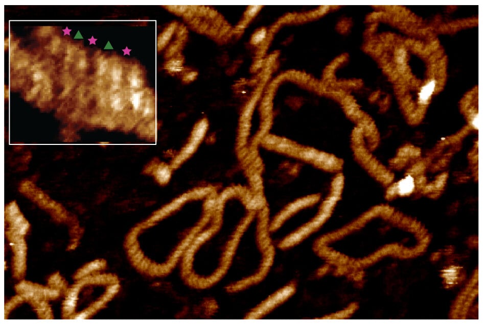
Each molecule examined was already a double helix, made of two strands wound around one another. The team studied how two of them associate alongside each other.
Using high-resolution atomic force microscopy, the researchers mapped the molecules' surfaces in liquid. The resulting images resolved the major and minor grooves spiraling around DNA, revealing instances of close alignment.
The images resolving individual grooves were obtained with nickel ions, which enabled optimal spatial resolution. Larger-area microscopy scans also examined pairing with magnesium and calcium, while simulations explored their effects on groove alignment.
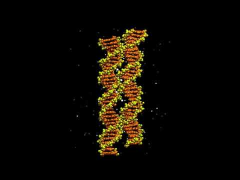
To show DNA progressively zipping together in a movie, simulations explored the movement behind the observed structures.
"The microscopy images were taken in static form, which allows us to obtain the resolution where we can observe and measure the individual minor and major grooves on each molecule," Sheffield biophysicist Alice Pyne told ScienceAlert.
That detail was essential, as simply seeing two molecules touch would not reveal whether their grooves lined up as the proposed zipper model predicted.
"The way two DNA duplexes zip together was hypothesized over 20 years ago," York biophysicist Agnes Noy told ScienceAlert.
"These images represent the first visualization that this idea is real."
The next question was what could keep the negatively charged molecules together.
"The ions help create a salt bridge between the two molecules, which holds them together," Pyne explained.
In simulations tracking individual atoms, ions carrying two positive charges connected neighboring helices. Some of the strongest contacts formed when their minor grooves aligned, allowing ions to interact with both molecules across the gap.
DNA's sequence influenced those connections, too.
"What the simulation showed is that there are special sequences that preferentially form these bridges, pinning the two molecules together, which then allows the molecules to 'zip' together," Pyne told ScienceAlert.
With nickel ions, particularly stable contacts were associated with a short DNA sequence called GTAC. In the simulations, magnesium and calcium also stabilized aligned pairings, although the networks of bridges differed between ion types.
This suggests the sequence helps shape where contacts form and how stable they become.
"In addition, we found that this DNA zipping depends on sequence and so genomes can present certain hot-spots where the pairing is especially easy," Noy says.
Understanding such preferences could help explain how matching DNA regions associate, a necessary step in genetic recombination. The new study offers a molecular framework for investigating that recognition, rather than demonstrating the entire process inside cells.
Indeed, the microscopy revealed several pairing arrangements. Most were incompatible with fully matching sequences aligning throughout the interacting region.
The researchers propose that initial attachment and extended pairing may involve different steps. Ion bridges could establish local contacts, while matching sequences help maintain alignment and allow pairing to extend farther along the helices.
So two molecules touching doesn't necessarily mean they have found a matching partner.
The experiments used purified DNA in controlled laboratory conditions. Inside cells, proteins and other molecules also help organize genetic material, including its wrapping around histone proteins to form structures called nucleosomes.
"These experiments show us how single molecules behave in a laboratory environment," Pyne told ScienceAlert.
She says the findings could help researchers understand DNA packaging beyond nucleosome wrapping, including how strongly negatively charged molecules pack so closely together.
Noy said investigating a possible connection between the findings and cancer will require further research.
The study didn't establish that these interactions cause cancer or map pairing hotspots across a genome. Identifying the sequences that define these hotspots, and locating them in the genome, is the next step.
For now, researchers have moved from a proposed arrangement to detailed images of aligned helices, supported by simulations revealing how ions could hold them together.
That gives them a concrete mechanism to test in the more complex environment of a cell.
The research has been published in Nucleic Acids Research.
This article was fact-checked by Rebecca Dyer and edited by Rebecca Dyer. While we pride ourselves on our process, we are only human. If you spot a mistake, please let us know.