As the COVID-19 pandemic crests wave after wave in many parts of the world, researchers have delivered a new look at the tiny coronavirus responsible for the huge chaos.
Each SARS-CoV-2 virus particle - virion - is a spiked protein ball encasing genetic material, around 50-200 nanometres wide. Somehow, even a brightly false-coloured micrograph infecting human lung cells can make them look almost… ornamental.
Striking new images of the SARS-CoV-2 virus by biochemist Camille Ehre from the University of North Carolina (UNC) School of Medicine have been published in the New England Journal of Medicine; they reveal just how well these microscopic virions can infect our airways.
The team introduced SARS-CoV-2 into a lab culture of human bronchial epithelial cells – the cells that provide a barrier between the air in the lungs and our blood stream. They left the two to mingle for 96 hours, and then used a scanning electron microscope to record the results.
"Virus production was approximately 3×10⁶ plaque-forming units per culture, a finding that is consistent with a high number of virions produced and released per cell," the team writes.
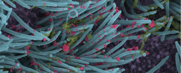
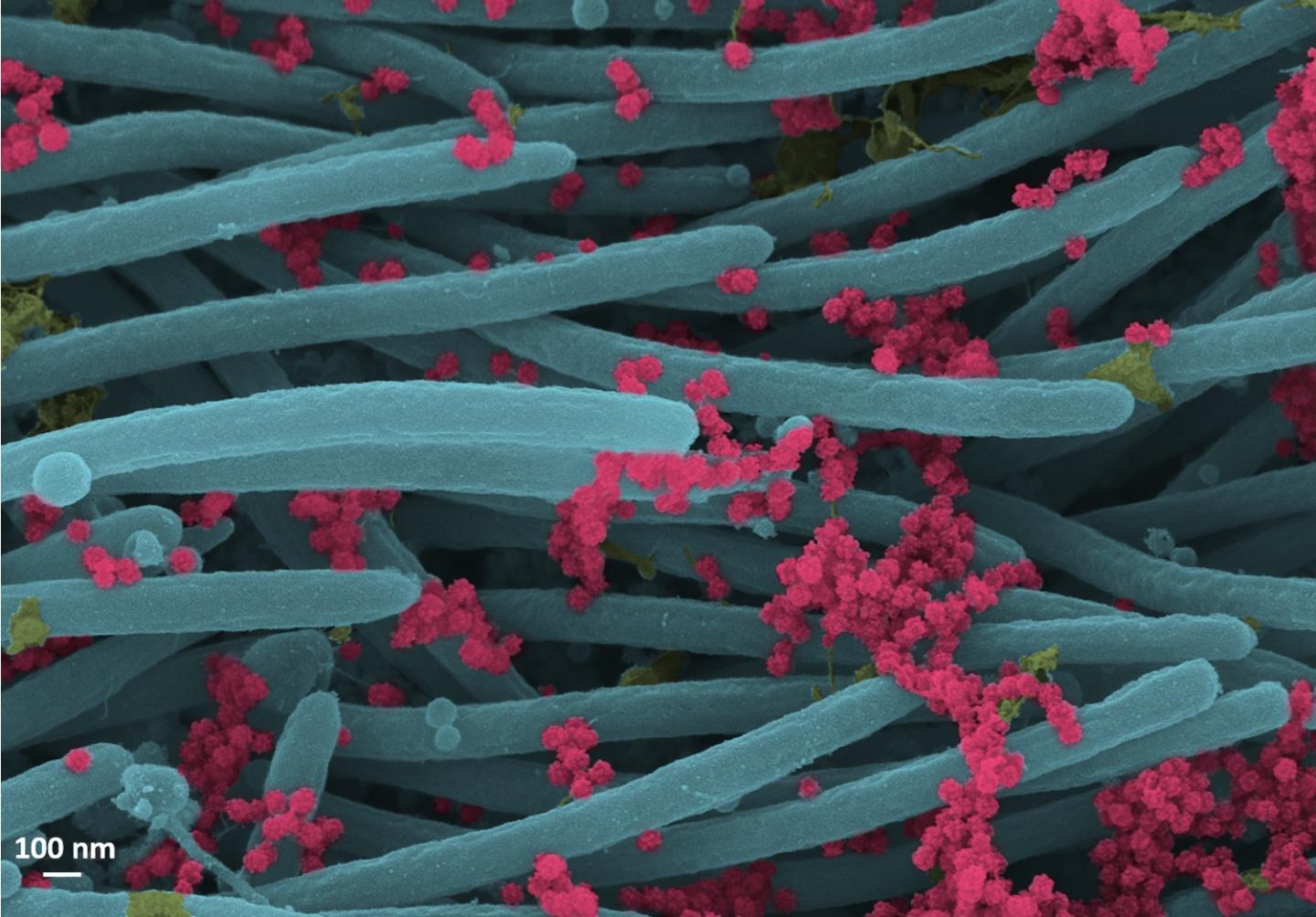
The images above and below have been falsely coloured by Cameron Morrison, a UNC medical student, and show hair-like structures called cilia in blue, strands of mucus in yellowish green, and SARS-CoV-2 in red.
The image at the top of this article shows the structures in a wider context, with small red virus particles, while the image below zooms in even more, clearly revealing the structure of the tiny particles and the density of the virus.
 (Ehre Lab/UNC School of Medicine)
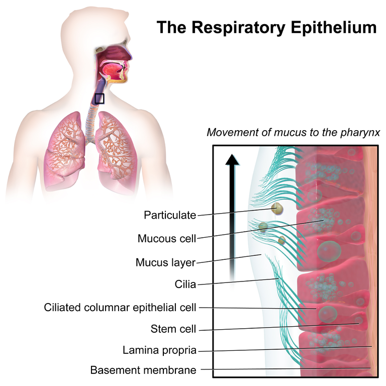
You might know of the cilia that reside in your gut, but they also exist inside the lungs, helping to protect our breathing organs from pathogens, keeping the lungs moist, and self-cleaning.
You can see an example of where they're found in the image below:
 (Blausen.com staff/WikiJournal of Medicine/CC BY 3.0)
Although the new micrographs really do look striking, they're more than just a novelty.
The images show the large number of virions produced when the virus hijacks our cells. In humans, the resulting virons are then likely coughed, talked, or even breathed out into the air in droplets, giving us a great reminder for people to wear masks, and keep your distance from others to limit the spread.
And maybe, looking at those tiny virus particles, we'll be able to better see the bigger picture.