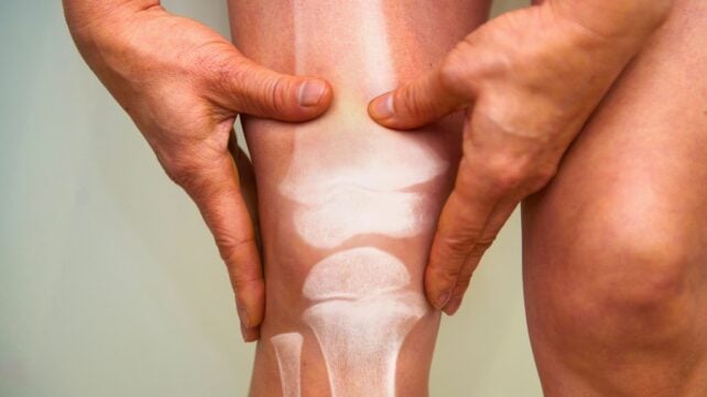
As time ticks on in our lives, various inevitable trends are noticeable, including our bones getting weaker and our brains getting slower.
But could one actually be an indicator of the other?
Researchers led by a team from Johns Hopkins University (JHU) in the US looked at the relationship between low bone mineral density (BMD) in the spine, and signs of cognitive decline, including two markers of brain damage that show up on scans.
The results showed, as reported in a study in Radiology, an association between weaker bones and signs of faster brain aging, including changes in white matter structure and a faster rate of cognitive decline.
"This study is the first longitudinal secondary analysis linking baseline vertebral bone mineral density to changes in white matter structure, white matter hyperintensity progression and cognition," says radiologist Shadpour Demehri, from JHU.
"This comprehensive study using both imaging and clinical assessments sends a clear signal that bone density at baseline is associated with both functional and imaging measures of age-related brain degeneration."
The study involved 715 participants in total, whose data had already been collected as part of a long-running health research project. As well as chest scans, these individuals had taken thinking and memory tests, which were followed up with brain scans and further cognitive tests several years after the first batch.
To get BMD readings from the chest CT scans, the researchers deployed a specially developed AI algorithm. Bone density refers to the presence of minerals such as calcium – if there's less of them present in the bone, it becomes weaker and more likely to break.
"Diagnostic images contain an immense amount of data that AI can now synthesize at scale across multiple organ systems," says Demehri.
"This creates a robust, newly feasible opportunity to interconnect co-existing age-related pathologies that have traditionally been studied in isolation and to identify shared biological pathways and potential common causal mechanisms."
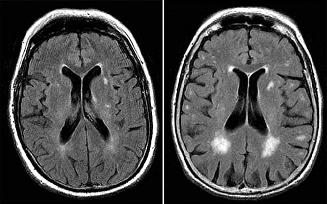
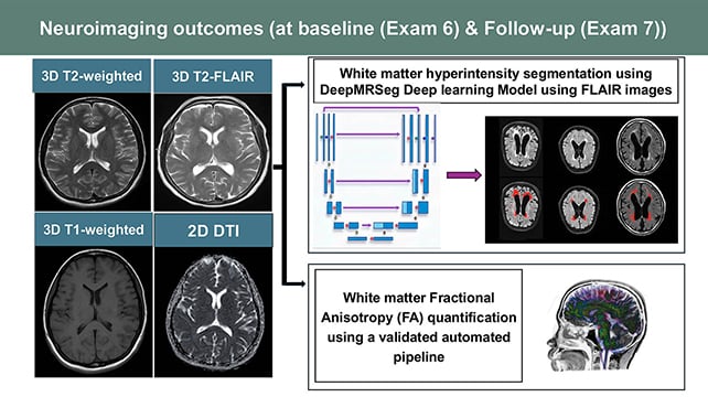
In some brain regions, lower BMD was linked to more white matter hyperintensities, which show up as bright spots on scans and highlight damaged tissue.
Lower BMD was also associated with white matter fractional anisotropy in other areas – an indicator of weaker brain structure.
The parts of the brain where these signs of damage were spotted have previously been linked to executive functioning (overall organization), working memory, and attention.
Faster decline in thinking test scores correlated to lower BMD as well.
The researchers are keen to emphasize that there's not enough evidence here to suggest weaker bones cause cognitive decline, and it's more likely that there's another factor involved driving both.
"This study is not about cause and effect, but rather the observation of a metabolic syndrome that may cause both bone and brain degeneration," says Demehri.
"The co-occurrence observed in the study may reflect shared metabolic drivers of aging, including insulin resistance, dyslipidemia, and menopausal change, rather than a direct bone-to-brain effect."
That individuals were followed over several years is to the study's credit, but longer-term research will be needed to see if the faster brain aging noticed here actually develops into diagnosed health issues such as dementia.
One of the ways this data could be used is to screen for people at higher risk of cognitive decline and related conditions, based on their BMD readings. However, much more data will need to be gathered and analyzed before predictions like that can be made.
What the research also shows is that with a little extra training and AI-powered reviewing, vast troves of existing medical data can be tapped into to reveal further insights.
"Chest CT is done for lung cancer screening, calcium scoring, nodule follow-up, and many other indications," says radiologist Sara Momtazmanesh, from JHU.
"These scans could provide an opportunistic measurement of bone density from the thoracic spine that is associated with a loss of cognitive function."
The research has been published in Radiology.