Researchers have managed to keep tabs on 1 million different neurons in the brains of mice at one time – taking scientists an impressive step closer towards one day being able to track the very-complex activity of human brains.
The key is an innovation that's being called 'light beads microscopy'. It improves on current two-photon microscopy, using lasers to trigger introduced fluorescence in living cells. As the cells are lit up, scientists can see how they're moving and interacting.
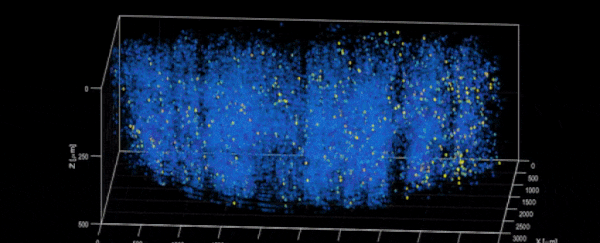
With light beads microscopy, scientists can get the speed, scale, and resolution required to map a mouse brain in detail as its neural activity changes. The near-simultaneous tracking can last for as long as the light beads are able to stay illuminated.
"Understanding the nature of the brain's densely interconnected network requires developing novel imaging techniques that can capture the activity of neurons across vastly separated brain regions at high speed and single-cell resolution," says neuroscientist Alipasha Vaziri, from the Rockefeller University in New York.
"We need to capture many neurons at distant parts of the brain at the same time at high resolution. These parameters are almost mutually exclusive."
In other words, current microscopy techniques have to choose between zooming in to get enough detail and missing out on everything that's going on, or zooming out to see the whole picture and losing some of the finer details.
Light beads microscopy is able to overcome these limitations by removing the dead time between laser pulses – using a cavity of mirrors it splits each single strong pulse into 30 smaller sub pulses of different strengths, which are then all able to reach different depths while keeping the same level of fluorescence.
This means multiple depths can be visualized in the same pulse, giving scientists a deeper, faster look at what's happening. The scientists have now demonstrated the technique to track 1 million neurons at once in a mouse brain.
"There's no good reason why we didn't do this five years ago," says Vaziri. "It would have been possible – the microscope and laser technology existed. No one thought of it."
Through light beads microscopy, the scientists are hoping to be able to track the interactions between the sensory, motor, and visual regions of the brain – not just in mice but in other animals too.
Interpreting and understanding the neural activity that's being recorded will require another step forward, but the new study pushes forward the idea of what's possible with this kind of microscopic analysis.
The better we can see inside the brain, the better we can work out how it operates – whether that's the interplay between individual nerve cells or finding out which parts of the brain correspond to which feelings and emotions.
"Light beads microscopy will allow us to investigate biological questions in a way that had not been possible before," says Vaziri.
The research has been published in Nature Methods.